Download
1 / 18
250 likes | 499 Vues
Physiology of the cochlea. Mechanical response of cochlea in response to sound Two major functions: Analysis of sound into components: Frequency/Spectral analysis Transduction of sound: Converting mechanical energy into electrochemical/neural energy.

E N D
Physiology of the cochlea Mechanical response of cochlea in response to sound Two major functions: • Analysis of sound into components: Frequency/Spectral analysis • Transduction of sound: Converting mechanical energy into electrochemical/neural energy http://www.neurophys.wisc.edu/animations/
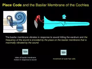
Mechanical response of cochlea to sound • Basilar membrane moves in response to stapes vibration • This vibration takes the form of a ‘traveling wave’ (von Bekesy) • Wave motion starts at the base and moves toward the apex http://www.iurc.montp.inserm.fr/cric/audition/english/corti/fcorti.htm
Instantaneous basilar membrane patternandenvelope of traveling wave
Characteristics of traveling wave • Points of maximum displacement: Peak • Peak depends on the frequency of incoming sound • Each part of basilar membrane ‘tuned’ to a particular frequency called critical/center/characteristic frequency (CF)
Low frequencies: Localized toward the apexHigh frequencies: Localized toward the base
Cochlea acts as a series of bandpass filters Incoming sound broken down into individual sinusoidal components Basilar membrane vibrates in response to each of these components Frequency/Spectral analysis
Basilar membrane mechanics • Greater the stimulus level, greater the amount of basilar membrane displacement • Temporal pattern of basilar membrane vibration follows that of incoming sound
Input-output functions • At mid-intensity levels, basilar membrane vibration increases with intensity in a non-linear or compressive fashion • At very low (< 30 dB SPL) and very high (> 90 db SPL), linear vibration • This is frequency dependent: Only for the CF • Above and below the CF: Linear input-output functions
Steps in transduction Mechanical vibrations translated into neural responses in the auditory nerve Stapes vibration sets inner ear fluid into vibration Basilar membrane vibrates Shearing motion of tectorial membrane Stereocilia of haircells bend http://www.iurc.montp.inserm.fr/cric/audition/english/corti/fcorti.htm
Bending of stereocilia http://www.iurc.montp.inserm.fr/cric/audition/english/corti/hcells/ohc/fohc.htm http://www.iurc.montp.inserm.fr/cric/audition/english/corti/fcorti.htm
Transduction, Cont’d. Increase in number of open ion channels at tip links Influx of positive pottassium and calcium ions into the cell body Resting membrane potential of endolymph: + 80 mv Intracellular resting potential: OHC: -70mv IHC: - 45mv Change in membrane potential Release of neurotransmitter Neuron at the base of the haircell fires
Transduction, Cont’d. Increase in number of open ion channels at tip links Influx of positive pottassium and calcium ions into the cell body Resting membrane potential of endolymph: + 80 mv Intracellular resting potential: OHC: -70mv IHC: - 45mv Change in membrane potential Release of neurotransmitter Neuron at the base of the haircell fires
Differences between OHC and IHC IHC: True sensory part of the cochlea Only forward transduction (Mechanical to neural) OHC: Both sensory and motor functions Forward and backward transduction (Mechanical to neural, and neural to mechanical)
Physiology of OHC and IHC OHC display ‘motility’ (change in length) in response to stimulation. IHC do not. Because of muscular contractions within the cell body and efferent connections to the OHC bodies. Thought to be basis of ‘cochlear amplifier’ and ‘active cochlea’
What is the cochlear amplifier? ‘Feedback loop’ within the Organ of Corti, because of OHC motility. Forward transduction: Basilar membrane moves, stereocilia are deflected, tranduction current flows into the OHC. This transduction current causes a change in the receptor potential (depolarization). Reverse transduction: Depolarization triggers the motility of the OHC. This change in length exerts force on the basilar membrane, deflecting the stereocilia and so on. OHC feed energy back into the basilar membrane!!
Evidence for active versus passive cochlea History: von Bekesy measured broad traveling waves in dead cochleae. However, finely-tuned tuning curves measured for auditory neurons. Fine tuning of basilar membrane is lost when OHC die. Compressive non-linearity (as seen in input-output curve) lost when OHC are damaged. Most important evidence for active cochlea: Oto-acoustic emissions.
Otoacoustic emissions (OAE) Responses that originate within the inner ear May be spontaneous or evoked Generated due to The feedback system in the cochlea Active processes within the cochlea Non-linearities within the cochlea
Tuning in the basilar membrane Vibration of basilar membrane can be described by a ‘tuning curve’ Amplitude required for the basilar membrane to vibrate at a certain constant displacement, as a function of the frequency of the input sound