Download
1 / 24
250 likes | 605 Vues
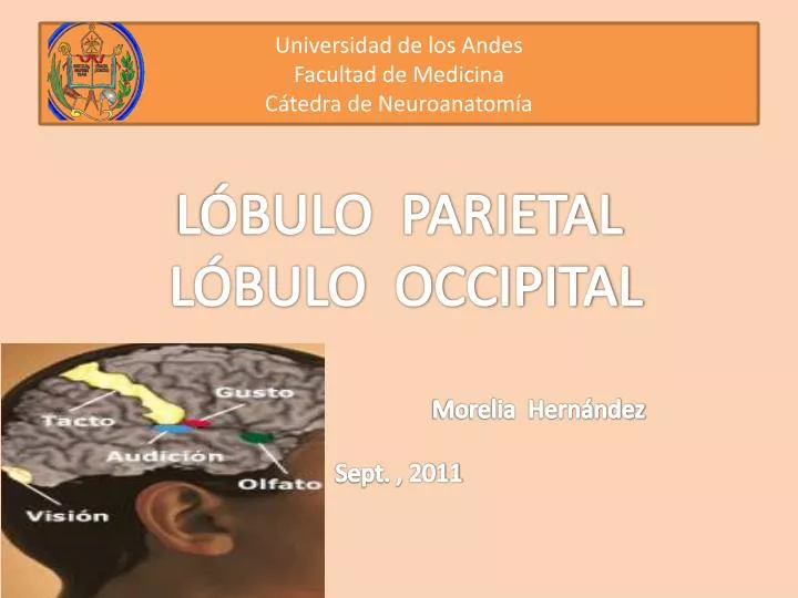
Universidad de los Andes Facultad de Medicina Cátedra de Neuroanatomía. LÓBULO PARIETAL LÓBULO OCCIPITAL Morelia Hernández Sept. , 2011. LÒBULO PARIETAL LÒBULO OCCIPITAL. HISTORIA. ALBERT EINSTEIN . Thomas Stolz Harvey(EEUU ) Cels gliales Sandra Witelson

E N D
Universidad de los AndesFacultad de MedicinaCátedra de Neuroanatomía LÓBULO PARIETAL LÓBULO OCCIPITAL Morelia Hernández Sept. , 2011
HISTORIA ALBERT EINSTEIN Thomas Stolz Harvey(EEUU ) Celsgliales Sandra Witelson (canadà) PESO 1400 Grs LÒBULO PARIETAL ANCHO
CITOARQUITECTURA LÓBULO PARIETAL KORBINIAN BRODMAN 1868-1918 AREAS FUNCIONALES DEL CEREBRO Estimulación Eléctrica : Lóbulo Occipital Alucinaciones Visuales . Complicada trama de Fibras Tálamo Mismo Hemisferio Otro Hemisferio
ÀREAS DE BRODMAN.LÒBULO PARIETAL • ÀREAS 3, 1 y 2 • PRIMARIAS • ÀREAS 5 Y 7 DE ASOCIACIÒN • CONSCIENCIA DEL ESQUEMA CORPORAL • RECONOCER OBJETOS • ÀREA 39 RECONOCIMIENTO ESPACIAL • ÀREA 40 COMPRENSIÒN LENGUAJE ORAL Y ESCRITO
AFERENCIAS DEL ÁREA SOMATO-SENSORIAL PRIMARIA • Cortico-Corticales TALAMO • Tálamo-Corticales • Colinérgicas
EFERENCIAS DEL ÀREA SOMATOSENSORIAL PRIMARIA • CORTICO-TALÀMICAS • CORTEZA-NUCLEOS COLINÈRGICOS • CORTEZA-NÙCLEOS NORADRENÈRGICOS • CORTICO-HIPOTALÀMICAS
AFERENCIAS DE LA CORTEZA DE ASOCIACIÓN • Recibe información del Área Somato-sensoriaL • TÀLAMO (PULVINAR) • FIBRAS DE ASOCIACIÒN • FIBRAS COMISURALES
LESIONES DE LÒBULO PARIETAL AGNOSIA TACTÌL ASTEREOGNOSIA CONTRALATERAL NEGACIÒN CORTICAL APRAXIA ALTERACION MOVIMIENTOS CUERPO Y DEL TACTO FINO
LÒBULO OCCIPITAL CORTE VERTICAL SUPERFICIE MEDIAL -Capa 4 (Bandas de Baillanger). Área Estriada LINEAS DE GENNARI
ÁREA VISUAL PRIMARIA CORTEZA VISUAL PRIMARIA UBICACIÓN: Superficie medial de lobulo occipital CENTRO DE LA RETINA:Pared posterior área 17. RETINA PERIFÉRICA (Anterior del área 17) Rodea Surco Calcarino Entre el cuneo y la cisura calcarina
corteza visual Imagen mental captada por el cerebro. Percepción colores formas relaciones espaciales movimientos.
AFERENCIASLÒBULO OCCIPITAL TÁLAMO ÁREA 17 NÚCLEO GENICULADO LATERAL (Geniculo-Calcarina) ÀREA 19 NUCLEO PULVINAR (ÁREA PREOCCIPITAL)
EFERENCIAS DEL ÀREA VISUAL PRIMARIA • CORTICO-TALÁMICAS • CORTEZA-NÚCLEOS COLINÉRGICOS • CORTEZA-NÚCLEOS NORADRENÉRGICOS • CORTICO-HIPOTALÁMICAS
EFERENCIAS DEL ÀREA VISUAL PRIMARIA • CORTICO-TALÀMICAS • CORTEZA-NUCLEOS COLINÈRGICOS • CORTEZA-NÙCLEOS NORADRENÈRGICOS • CORTICO-HIPOTALÀMICAS
Lesiones del lòbulo Occipital • Amaurosis • Daño es contralateral • Agnosia Visual • Acromatopsia • Prosopagnosia • Hemianopsia
CASO CLÌNICO • Paciente masculino de 45 años de edad natural del Táchira y procedente de Barinas, quien inicia enfermedad actual hace 4 meses con cefalea, disminución de fuerza muscular y episodio convulsivo , fue dado de alta con tratamiento anticomicial, 4 meses después acude con cefalea fronto-occipital tipo opresivo y disminución progresiva de la fuerza muscular. • RMN: Evidencia imagen para sagital parietal derecha, heterogénea, híper intensa en t1 y t2, con áreas hipo intensas en su interior que impresiona área de necrosis, con moderado edema perilesional y escaso efecto de masa. Se realizó exéresis tumoral en un 80%. La biopsia reporto Glioblastoma Multiforme. Actualmente se encuentra en terapia física
CONCLUSIONES • LÓBULO PARIETAL: SOMATOSENSORIAL PRIMARIA • (ÀREAS 3, 1 Y 2 BRODMAN) • CORTEZA DE ASOCIACIÓN (ÁREAS 5, 7, 39, 40) • SENSACIONES. (LENGUAJE,TACTO). • ORIENTACIÓN ESPACIAL (Segmentos corporales). • LÓBULO OCCIPITAL • CORTEZA VISUAL PRIMARIA (ÁREA 17 ) • (ADYACENCIAS LÒBULO TEMPORAL • Y PARIETAL). • ASOCIACIÓN ÁREAS 18 Y 19 BRODMAN
BIBLIOGRAFÌA • 1.-AFIFI,A.BERGMAN,R.2006.NEUROANATOMÍA FUNCIONAL.MAC GRAW-HILL.INTERAMERICANA. • 2.- BARR, K.2006.EL SISTEMA NERVIOSO HUMANO.UN PUNTO DE VISTA ANATÒMICO.MCGRAW-HILL.INTERAMERICANA. • 3.-SNELL,R.2008.NEUROANATOMÍA CLÍNICA.EDITORIAL PANAMERICANA.