Download
1 / 27
310 likes | 1.04k Vues
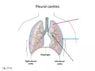
Management of spontaneous pneumothorax : British Thoracic Society pleural disease guideline 2010. EM/CS R2 박시열. Pneumothorax. Air in the pleural cavity Secondary pneumothorax (SSP) With underlying lung disease Primary pneumothorax (PSP)

E N D
Management of spontaneous pneumothorax: BritishThoracic Society pleural disease guideline 2010 EM/CS R2 박시열
Pneumothorax • Air in the pleural cavity • Secondary pneumothorax (SSP) • With underlying lung disease • Primary pneumothorax(PSP) • Subpleural blebs and bullae was found upto 90% • SSP is associated with a higher morbidity and mortality than PSP
Clinical evaluation • Symptoms in PSP may be minimal or absent. • Symptoms are greater in SSP, even if the pneumothorax is relatively small in size • The presence of breathlessness influences the management strategy • Severe symptoms and signs of respiratory distress suggest the presence of tension pneumothorax
Imaging • Diagnosis of pneumothorax is usually confirmed by imaging techniques • Clinical evaluation should be the main determinant of the management strategy • Standard erect chest x-rays in inspiration are recommended • Expiratory films are not thought to confer additional benefit
Imaging • 1. Standard erect PA chest x-ray. • 2. Lateral x-rays. • 3. Expiratory films. • 4. Supine and lateral decubitus x-rays. • 5. Ultrasound scanning. • 6. Digital imaging. • 7. CT scanning.
Size of pneumothorax • Less important than the degree of clinical compromise • Plain PA chest x-ray has limitation • 2 cm radiographic pneumothorax approximates to a 50% by volume • CT scanning is the best tool for establishing the size of a pneumothorax
Treatment • Distinguish PSP vs SSP • Breathlessness indicates the need for active intervention • Consider size of pneumothorax
Treatment • Observation is the treatment of choice for small PSP without significant breathlessness • Selected asymptomatic patients with a large PSP may be managed by observation alone • Patients with a small PSP without breathlessness should be considered for discharge with early outpatient review
Treatment • Chest drain insertion, others • Tension pneumothorax and bilateral pneumothorax • Hydropneumothorax
Treatment • Needle aspiration or chest drain • Needle (14e16 G) aspiration (NA) is as effective as large-bore (>20 F) chest drains • Associated with reduced hospitalisation and length of stay • NA should not be repeated unless there were technical difficulties • Following failed NA, small-bore (<14 F) chest drain insertion is recommended
Treatment • NA should cease after 2.5 l of air has been aspirated • likely presence of a persistent air leak
Treatment • Suction • Suction should not be routinely employed • Caution is required because of the risk of RPO • High-volume low-pressure suction systems are recommended
Management of SSP • All patients with SSP should be admitted at least 24 h and receive O2 • Most patients will require small-bore chest drain • All patients will require early referral to a chest physician • Persistent air leak should be discussed with a thoracic surgeon at 48 h
Management of SSP • SSP but unfit for surgery • Medical • Ambulatory management with a Heimlich valvepleurodesis may be appropriate
Discharge & follow up • Return to hospital if increasing breathlessness develops • Followed up by respiratory physicians until full resolution • Air travel should be avoided until full resolution
Medical chemical pleurodesis • Control difficult or recurrent pneumothoraces • surgical options are more effective • Performed by a respiratory specialist
Referral to thoracic surgeon • Second ipsilateralpneumothorax. • First contralateralpneumothorax. • Synchronous bilateral spontaneous pneumothorax. • Persistent air leak (despite 5~7 days of chest tube drainage) or failure of lung re-expansion. • Spontaneous haemothorax • Professions at risk (eg, pilots, divers) • Pregnancy
Surgical strategies • Open thoracotomy and pleurectomy • lowest recurrence rate (approximately 1%) for difficult or recurrent pneumothoraces • Video-assisted thoracoscopic surgery (VATS) with pleurectomyand pleural abrasion is better tolerated but has a higher recurrence rate of approximately 5%.
Surgical strategies • Surgical chemical pleurodesis • 5 g sterile graded talc, with which the • complications • ARDS and empyemaare rare
Tension pneumothorax • Medical emergency • Requires heightened awareness in a specific range of clinical situations • O2 and needle decompression • A standard cannula may be insufficiently long if used in the 2nd intercostalspace • Chest drain should be inserted immediately after needle decompression
Tension pneumothorax • 1. Ventilated patients on ICU. • 2. Trauma patients. • 3. Resuscitation patients (CPR). • 4. Lung disease, especially acute presentations of asthma and COPD • 5. Blocked, clamped or displaced chest drains. • 6. Patients receiving non-invasive ventilation (NIV). • 7. Miscellaneous group, for example patients undergoing hyperbaric oxygen treatment.
Pneumothorax & pregnancy • Recurrence is more common • Risks to the mother and fetus • Requires close cooperation between chest physicians, obstetricians and thoracic surgeons • Simple observation and aspiration are usually effective during pregnancy
Pneumothorax & pregnancy • Simple observation and aspiration are usually effective during pregnancy • Elective assisted delivery and regional anaesthesiaat or near term • If the mother is not dyspnoeic, no fetal distress and the pneumothorax is small (<2 cm) • Corrective surgical procedure (VATS) should be considered after delivery
Iatrogenic pneumothorax • Transthoracic needle aspiration (24%) • Subclavianvessel puncture (22%) • Thoracocentesis (22%) • Pleural biopsy (8%) • Mechanical ventilation (7%)
Iatrogenic pneumothorax • Resolve spontaneously by observation alone • Simple asp • Those on positive pressure ventilation require chest drain insertion as positive pressure maintains the air leakiration
Others • HIV infection • Requires early intercostal tube drainage and surgical referral, in addition to appropriate treatment for HIV and PJP infection. • Cystic fibrosis • Early and aggressive treatment with early surgical referral • Pleural procedures, including pleurodesis, do not have a significant adverse effect on the outcome of subsequent lung transplantation