Download

1 / 63

E N D
Dr. Luca Alina Hypertension
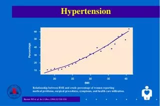
Hypertension is a major cause of morbidity and mortality in the United States and in many other countries. The prevalence of hypertension in the United States for people aged 60-69 years is more than 50%. This prevalence increases to approximately 75% among those aged 75 years. Hypertension is now commonly discovered in children. The long-term health risks to these children with hypertension may be substantial
The Fourth Report introduced a new category called prehypertension. The condition is diagnosed when a child's average BP exceeds the 90th percentile but is less than the 95th percentile. Any adolescent whose BP is greater than 120/80 mm Hg is also given this diagnosis, even if their reading is less than the 90th percentile. This classification was created to align the categories for children with the categories for adults from the 2003 recommendations of the Seventh Report of the Joint National Committee on Prevention, Detection, Evaluation, and Treatment of High Blood Pressure.
Stage I hypertension is diagnosed if a child's BP is greater than the 95th percentile but less than or equal to the 99th percentile plus 5 mm Hg. A child is classified as having stage II hypertension if their BP is greater than the 99th percentile plus 5 mm Hg. • If the systolic and diastolic classifications cause a discrepancy, the child's condition should be categorized by using the higher value. Table 1 serves as a guide to the practicing physician. Full tables from the NHLBI may be found at Blood Pressure Tables for Children and Adolescents.
Hypertension can be primary (ie, essential) or secondary. • In general, the younger the child and the higher the BP, the greater the likelihood that hypertension is secondary to an identifiable cause. A secondary cause of hypertension is most likely to found before puberty. After puberty, hypertension is likely to be essential. • A review of literature revealed that 78% of 563 young patients with secondary hypertension had a renal parenchymal abnormality. In the remaining 22%, the cause of hypertension, in order of frequency, was renal artery stenosis, coarctation of the aorta, pheochromocytoma, and a variety of other conditions. Causes
BP is determined by the balance between cardiac output and vascular resistance. A rise in either of these variables, in the absence of a compensatory decrease in the other, increases mean BP, which is the driving pressure. Several factors regulate cardiac output and vascular resistance. Pathophysiology
Factors that affect BP include the following: • Cardiac output • Baroreceptors • Extracellular volume • Effective circulating volume • Atrialnatriuretic hormones • Mineral corticoids • Angiotensin • Sympathetic nervous syndrome Factors
Vascular resistance • Pressors • Angiotensin II • Calcium (intracellular) • Catecholamines • Sympathetic nervous system • Vasopressin • Depressors • Atrialnatriuretic hormones • Endothelial relaxing factors • Kinins • Prostaglandin E 2 • Prostaglandin I 2
United States The true incidence of hypertension in the pediatric population is not known. This vagueness partly stems from the somewhat arbitrary definition of hypertension. In adults, hypertension is defined on the basis of data from extensive studies that allowed for correlation of BP with adverse events, such as heartfailure or stroke. Frequency
Similar studies have not been performed in children, although reports from small populations of children provided compelling evidence of a relationship between hypertension and both ventricular hypertrophy and atherosclerosis. In children, the definition of hypertension is based exclusively on frequency-distribution curves for BP. As a consequence, estimations of the prevalence of pediatric hypertension lack a scientific basis. The number of children who might be defined as having hypertension and the frequency with which they develop complications during adulthood remains unknown.
International Because of differences in genetic and environmental factors, incidences vary from country to country and even from region to region in the same country.
Race • The Task Force on Blood Pressure Control in Children noted no differences in BP between African American and Caucasian children. However, peripheral vascular resistance and sensitivity of BP to salt intake appear greater in African American children than in Caucasian children, at any age. Sex • No significant differences are observed in BP between girls and boys younger than 6 years. From that age until puberty, BP is slightly higher in girls than in boys. At puberty and beyond, BP is slightly higher in male adolescents and men than in comparably aged female adolescents and women.
Age Height and weight affect BP. However, these relationships do not become evident until children are school aged. The Task Force on Blood Pressure Control in Children considered these factors when they published their normative data in 1987. Numerous investigators have noted a correlation between the BP of parents and that of their offspring. Familial aggregation of BP is detectable early in life. Some data relate this association to concomitant obesity in both parent and child.
A well-taken history provides clues about the cause of hypertension and guides the nature and sequence of ensuing investigations. ClinicalHistory
Relevant information includes the following: • Prematurity • Bronchopulmonary dysplasia • History of umbilical artery catheterization • Failure to thrive • History of head or abdominal trauma • Family history of heritable diseases (eg, neurofibromatosis, hypertension) • Medications (eg, pressor substances, steroids, tricyclic antidepressants, cold remedies, medications for attentiondeficit hyperactivity disorder [ADHD]) • Episodes of pyelonephritis (perhaps suggested by unexplained fevers) that may result in renal scarring • Dietary history, including caffeine, licorice, and salt consumption • Sleep history, especially snoring history • Habits, such as smoking, drinking alcohol, and ingesting illicit substances
Presenting symptoms and signs are not specific in neonates and absent in most older children unless their hypertension is severe.
Signs and symptoms that should alert the physician to the possibility of hypertension include the following: • Neonates • Failure to thrive • Seizure • Irritability or lethargy • Respiratory distress • Congestive heart failure • Children (Findings in addition to those observed in neonates) • Headache • Fatigue • Blurred vision • Epistaxis • Bell palsy
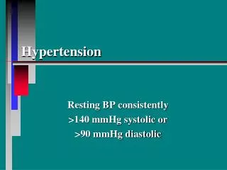
Measurement and recording of blood pressure (BP) • Best medical care includes yearly measurement of BP in every child older than 3 years, preferably by means of auscultation with a mercury gravity manometer. Doppler and oscillometric techniques can be used in children in whom auscultatory BP measurements are difficult to obtain. Measurements obtained by using oscillometric devices that exceed the 90th percentile should be repeated with auscultation. Physical
Measurements repeated over time are required to obtain meaningful information. • Proper cuff size is essential for accurate measurement of BP. The width of the rubber bladder inside the cloth cover should cover at least 40% of the patient's arm circumference at a point midway between the olecranon and the acromion. The length of the bladder in the cuff should cover 80-100% of the circumference of the arm. If a cuff is too small, the next larger cuff size should be used, even if it appears too large.
The child should be relaxed and in a comfortable, preferably sitting, position with the child's feet on the floor and the back supported. The patient's right arm should be resting on a supportive surface at the level of the heart. Infants can be examined while supine. • The cuff should be inflated at a pressure approximately 20 mm greater than that at which the radial pulse disappears and then allowed to deflate at a rate of 2-3 mm Hg/s.
The first Korotkoff sound (ie, appearance of a clear tapping sound) defines the systolic pressure, whereas the fifth Korotkoff sound (ie, disappearance of all sounds) defines the diastolic pressure. The fourth (low-pitched, muffled) sound and the fifth sound frequently occur simultaneously, or the fifth sound may not occur at all. Diastolic BP must be recorded. When Korotkoff sounds can be heard down to 0 mm Hg, the BP measurement should be repeated with less pressure applied to the head of the stethoscope than was applied before.
Systolic BP in the lower extremities must be measured when elevated systolic BP in the upper extremities is first noted regardless of whether amplitude of the arterial pulse seems lower in the legs to be lower than that in the arms. Increased systolic pressure in the arm suggests coarctation of the aorta. If found, systolic pressure must also be measured in the left arm and leg. With the patient in the supine position, place a cuff on the calf. The cuff should be wide enough to cover at least two thirds of the distance from knee to ankle. Doppler sonography can be used to detect onset of blood flow, which reflects systolic BP, in the posterior tibial or dorsalispedis artery. The value should be compared with a similarly obtained Doppler systolic BP in the arm, again with the patient supine.
Remember that the artifact of distal pulse amplification causes the measured systolic BP at the brachial artery to be less than that at the posterior tibial or dorsalispedis artery. This difference may be only a few millimeters in the infant but can rise to 10-20 mm Hg in the older child or adult. Magnitude of this artifact is directly proportional to the pulse pressure. In a patient with chronic aortic regurgitation, for example, the difference in measured systolic pressure may exceed 40 mm Hg. At no time should the systolic pressure in the arm exceed that in the foot. If it does, pressures in both arms and legs should be measured. Consistent recording of high arm systolic pressure indicates aortic coarctation. High pressure in only the right arm suggests that an obstruction is present proximal to origin of the left subclavian artery.
Interpretation of BPs • Hypertension is defined as average systolic or diastolic BPs greater than those at the 95th percentile. Any child with a BP exceeding the 90th percentile requires scrutiny. • Patients with severe hypertension and target-organ damage require immediate attention. For other patients, several measurements of BP should be made at weekly intervals to determine if the elevation is sustained. • The average of multiple measurements should be plotted on an appropriate percentile chart. If the average measurement is between the 90th and 95th percentiles (ie, prehypertensive) the child's BP should be monitored at 6-month intervals. If the average BP is greater than the 95th percentile, the child should be evaluated further and therapy considered.
Patients with stage I hypertension should be seen again in 1-2 weeks. Those with stage II hypertension should be reevaluated in 1 week or sooner if the patient is symptomatic. • White-coat hypertension is diagnosed in a patient who has a BP above the 95th percentile when measured in the physician's office but who is normotensive outside the clinical setting. Ambulatory monitoring of BP usually is required to diagnose white-coat hypertension.
Objective of physical examination: A primary objective of the physical examination is to identify signs of secondary hypertension, including the following: • Body mass index to assess for metabolic syndrome • Tachycardia to assess for hyperthyroidism, pheochromocytoma, and neuroblastoma • Growth retardation to assess for chronic renal failure • Café au lait spots to assess for neurofibromatosis • Abdominal mass to assess for Wilms tumor and polycystic kidney disease
Epigastric and/or abdominal bruit to assess for coarctation of the abdominal aorta or renal artery stenosis • BP difference between upper and lower extremities to assess for coarctation of the thoracic aorta • Thyromegaly to assess for hyperthyroidism • Virilization or ambiguity to assess for adrenal hyperplasia • Stigmata of Bardet-Biedl, von Hippel-Landau, Williams, or Turner syndromes • Acanthosis nigricans to assess for metabolic syndrome
Turner syndromes Lymphedema of the feet in an infant is shown Hairline and a shield-shaped chest. Hyperconvex nails in Turner syndrome
In patients with hypertension, proceed from simple tests that can be performed in an ambulatory setting to complex noninvasive tests and finally to invasive tests. Findings from the patient's history and physical examination dictate the appropriate order of tests. • On urine dipstick testing, a positive result for blood and/or protein indicates renal disease. • Urine cultures are used to evaluate the patient for chronic pyelonephritis. WorkupLaboratory Studies
The CBC count may indicate anemia due to chronic renal disease. Blood chemistry may be helpful. An increased serum creatinine concentration indicates renal disease. Hypokalemia suggests hyperaldosteronism. High urinary excretion of catecholamines and catecholamine metabolites (metanephrine) indicates pheochromocytoma or neuroblastoma. Fasting lipid panels and oral (PO) glucose-tolerance tests are performed to evaluate metabolic syndrome in obese children. Urine sodium levels reflect dietary sodium intake and may be used as a marker to follow up a patient after dietary changes are attempted.
Blood hormone levels may be measured. High plasma renin activity indicates renal vascular hypertension, including coarctation of the aorta, whereas low activity indicates glucocorticoid remediable aldosteronism, Liddle syndrome, or apparent mineralocorticoid excess. A high plasma aldosterone concentration is diagnostic of hyperaldosteronism. High values of catecholamine (epinephrine, norepinephrine, dopamine) are diagnostic of pheochromocytoma or neuroblastoma. Drug screening is performed to identify substances that might cause hypertension.
Echocardiography • Left ventricular hypertrophy results from chronic hypertension. This finding confirms the chronicity of the hypertension and is an absolute indication for starting or intensifying treatment. • Left ventricular hypertrophy is symmetric, consisting of equivalent increases in thickness of both the left ventricular portion of the ventricular septum and the left ventricular posterior wall. • Also assess left ventricular function. • Echocardiography is essential in the evaluation of suspected aortic coarctation. Precise anatomic detail of the aortic arch and its branches must be obtained. Imaging Studies
Abdominal ultrasonography • This test may reveal tumors or structural anomalies of the kidneys or renal vasculature. • Renal scarring suggests excessive renin release. • Asymmetry in renal size suggests renal dysplasia or renal artery stenosis. • Renal or extrarenal masses suggest a Wilms tumor or neuroblastoma, respectively. • Radionuclide imaging (without or with captopril): Asymmetry suggests renal artery stenosis. • Doppler studies: Asymmetry in renal artery blood flow suggests renal artery stenosis. • Digital subtraction arteriography: Asymmetry between the 2 renal arteries indicates renal artery stenosis.
Angiography • This test may reveal differences in the structure (diameter) of the renal vessels. • Sampling of blood from renal arteries, renal veins, and aorta may reveal differences in renin secretion between the kidneys. • A renin activity ratio of 3:1 between the kidneys is considered diagnostic of renal vascular hypertension. • Other tests • Cardiac catheterization is not necessary in the evaluation of aortic coarctation. • CT and MRI with angiography can provide further anatomic definition of an aortic coarctation, but neither study is necessary for diagnosis.
Nonpharmacologic measures • Nonpharmacologic measures are important in the treatment of all patients with hypertension, regardless of its etiology or severity. In children with mild or moderate hypertension, this approach may suffice to lower blood pressure (BP) to within normal limits. A nonpharmacologic approach avoids the need for drugs that have adverse effects and that require a degree of compliance difficult to achieve in children. Medical Care
Weight reduction should be a goal in overweight children with hypertension regardless of its etiology. Obesity and hypertension are closely correlated, particularly in adolescents. Aerobic and isotonic exercises have a direct beneficial effect on BP. They help in reducing excess weight or maintaining appropriate body weight. Encourage participation in sports. Only patients with severe uncontrolled hypertension or cardiac abnormalities that require exercise restriction are exempt from aerobic and isotonic exercises.
Salt restriction probably benefits only a subgroup of patients with hypertension, particularly African American patients, who may have a defect in the cellular handling of sodium. However, given the excessive amount of salt in the typical American diet, reduced salt intake should be recommended to all patients with hypertension. The Task Force recommends the Dietary Approaches to Stop Hypertension (DASH) eating plan Stress-reducing activities can reduce BP when performed on a regular basis. However, this effect is lost when the activity is discontinued.
Potassium supplementation can decrease BP and reduce ventricular hypertrophy in adults. How potassium supplementation affects children with hypertension remains to be tested. However, avoiding potassium depletion (eg, from diuretic therapy) and prescribing a potassium-rich diet in patients without renal insufficiency appear reasonable. When sleep-disordered breathing is discovered, weight loss, tonsillectomy and adenoidectomy, and/or use of continuous positive airway pressure may improve the patient's sleep and secondarily improve BP.
Management of hypertensive crisis • Hypertensive crises occur as a result of an acute illness, such as postinfectiousglomerulonephritis or acute renal failure, the excessive ingestion of drugs or psychogenic substances, or exacerbated moderate hypertension. • The clinical manifestations may be those of cerebral edema, seizures, heart failure, pulmonary edema, or renal failure. Remember that accurately assessing BP in every patient presenting with a seizure is essential, particularly when no seizure disorder has been established in that patient.