Download
1 / 75
840 likes | 2.1k Vues
Update on Chronic Venous Disease Treatment. By: Gabriel Bietz. Chronic venous disease. Most common vascular disorder 3 Billion US dollars spent a year for treatment 3 % of the total Heath care Budget 2 million USA work days lost per year. Definition.

E N D
Update on Chronic Venous Disease Treatment By: Gabriel Bietz
Chronic venous disease • Most common vascular disorder • 3 Billion US dollars spent a year for treatment • 3 % of the total Heath care Budget • 2 million USA work days lost per year
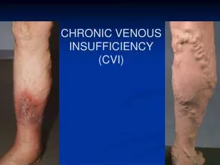
Definition Telangiectasias - are a confluence of dilated intradermal venules less than one millimeter in diameter. Reticular veins - are dilated bluish subdermal veins, one to three millimeters in diameter. Usually tortuous. Varicose veins - are subcutaneous dilated veins three millimeters or greater in size. They may involve the saphenous veins, saphenous tributaries, or nonsaphenous superficial leg veins.
Abnormal Veins Telangiectasias Varicose vein Reticular veins
Common Questions Are they dangerous? How do they form? Why does it happen? Did I inherit it? What tests can we use? What treatments are available?
Fascia, Veins and Cutaneous nerves of the LE The subcutaneous tissue of the hip & thigh is continuous with that of the inferior abdominal wall and buttock. At the knee the subcutaneous tissue loses its fat and blends with the deep fascia Deep fascia: strong & inelastic it invests the LE; it limits outward expansion of the contracting musculature, the increased pressure “pumps” the blood proximally through the veins.
Deep fascia of the thigh (fascia lata) The fascia lata attaches to and is continuous with: 1.The inguinal ligament, pubic arch, body of the pubis and pubic tubercle. 2.Scarpa’s fascia of the inferior abdominal wall attaches to deep LE fascia inferior to the inguinal ligament. 3. Iliac crest (lateral & posterior). 4. Sacrum, coccyx, sacrotuberous ligament, ischial tuberosity postreiorly. 5. Exposed parts of bones at the knee & deep fascia of the leg.
LE Compartments Anterior & Posterior intermuscular septa – pass from the deep crural fascia to attach to the margins of the fibula. Interosseous membrane traverses from tibia to fibula (4) compartments created. Anterior -dorsiflexors Lateral - fibular (everter) compartment Posterior – plantarflexor compartment The transverse intermuscular septum divides this into a deep & superficial compartment.
Crural fascia Deep fascia to the leg – continuous with the fascia lata, attaches to the anterior & medial borders of the tibia; it is continuous with the periosteum. Thinner distally but thickens to form an extensor/flexor retinaculum both anterior & posterior to the ankle.
Superficial veins Great saphenous – formed by the union of the dorsal digital vein of the great toe and the dorsal venous arch. Ascends anterior to the medial malleolus, posterior to the medial condyle of the femur. It freely communicates with the small saphenous vein. Proximally it traverses the saphenous opening in the fascia to enter the femoral vein.
Saphenous opening NAVEL This is a gap in the fascia lata infero-lateral to the inguinal ligament, lateral to the pubic tubercle. Medial margin is smooth Lateral margin is sharp forming the falciform ligament Cribiform fossa – a sleeve like membrane covering the saphenous opening
Small saphenous vein Formed by the union of the dorsal digital vein of the 5th digit and distal venous arch. Runs posterior to the lateral malleolus, lateral to the calcaneal tendon. Runs superiorly medial to the fibula and penetrates the deep fascia of the popliteal fossa, ascends between the heads of the gastrocnemius muscle to join the popliteal vein.
Perforating veins Penetrate the deep fascia, tributaries of the saphenous veins, valves are located just distal to penetration of the deep fascia. Veins cross the deep fascia obliquely Muscle contraction causes the valves to close prior to venous compression so blood is forced proximally (musculo-venous pump).
Deep Veins Usually paired with named arteries inside a vascular sheath, this allows arterial pulsation to force blood proximally. The popliteal vein joins the femoral vein in the popliteal fossa Femoral vein is joined by the deep vein of the thigh. The femoral vein passes deep to the inguinal ligament to become the external iliac vein.
Lymphatic drainage of LE Superficial Lymphatics Superficial lymphatic vessels accompany the saphenous veins (great & small) Superficial lymphatics end at the superficial inguinal nodes most of this lymph drains to the external iliac nodes, some drains to the deep inguinal nodes. Small lymphatics drain to the popliteal nodes Deep Lymphatics Deep lymphatics drain to the popliteal nodes which then drain to the inguinal nodes then to the external iliac nodes. Both deep & superficial drain into the lumbar lymphatics.
Varicose veins Varicose veins are a common condition in the United States, affecting up to 15 percent of men and up to 25 percent of women. For many people, varicose veins and spider veins a common, mild and medically insignificant variation of varicose veins — are simply a cosmetic concern. For other people, varicose veins can cause aching pain and discomfort. Sometimes the condition leads to more serious problems. Varicose veins may also signal a higher risk of other disorders of the circulatory system. Callam, MJ. Epidemiology of varicose veins. Br J Surg 1994;81:167.
Etiology Reflux 80% Venous obstruction 18-28% Resultant edema and skin changes = Postthrombotic syndrome Muscle Pump Dysfunction
Stasis Pathophysiology Usually associated with venous incompetence Primary and secondary reflux Edema Vein wall dilatation Inflammation/Pigmentation (Hemosiderin deposits) “Fibrin cuffing” Ulceration
Risk factors Age: Aging causes wear and tear. Eventually, that wear causes the valves to malfunction. Sex: Women > Men. Hormonal changes during pregnancy or menopause. Progesterone relaxes venous walls. HRT / OCP may increase the risk of varicose veins. Genetics Obesity: Increases venous HTN. Standing for long periods of time. Prolonged immobile standing impairs venous return. Fowkes, FG, Lee, AJ, Evans, CJ, et al. Lifestyle risk factors for lower limb venous reflux in the general population: Edinburgh Vein Study. Int J Epidemiol 2001; 30:846. Sadick, NS. Predisposing factors of varicose and telangiectatic leg veins. J Dermatol Surg Oncol 1992; 18:883. Iannuzzi, A, Panico, S, Ciardullo, AV, et al. Varicose veins of the lower limbs and venous capacitance in postmenopausal women: relationship with obesity. J Vasc Surg 2002; 36:965. Evans, CJ, Fowkes, FG, Hajivassiliou, CA, et al. Epidemiology of varicose veins. A review. Int Angiol 1994; 13:263.
Strong familial component Not well studied Twin studies 75% identical, 52% non identical If both parents VVS - 90% of children VVs If one parent was affected 25 percent for men and 62 percent for women Cornu-Thenard, A, Boivin, P, Baud, JM, et al. Importance of the familial factor in varicose disease. Clinical study of 134 families. J Dermatol Surg Oncol 1994; 20:318.
Symptoms Achy or heavy feeling, burning, throbbing, muscle cramping and swelling. Prolonged sitting or standing tends to intensify symptoms. Pruritis Painful skin ulcers
Complications Extremely painful ulcers may form on the skin near varicose veins, particularly near the ankles. Brownish pigmentation usually precedes the development of an ulcer. Occasionally, veins deep become enlarged. Bleeding Superficial thrombophlebitis
CEAP classification 1994 AVF Meeting
Patient Assessment History History of symptoms and onset History of venous complications Desire for treatment Comorbidities Rule out secondary cause including DVT and HEART Failure Examination Patient in general Pedal pulses Groins Veins Trendelenburg Test Venous claudication
Investigation All get a Duplex scan Examines – Deep veins – Superficial veins – Incompetence and patency Other Tests Physiologic testing Phlebography Intravascular Ultrasound
Duplex scan Vast majority have superficial incompetence only. Sensitivity 95 % for identifying the competence of the saphenofemoral and saphenopopliteal junctions. Less sensitive for identifying incompetent perforators (40 to 60 percent) Lin, JC, Iafrati, MD, O'Donnell, TF Jr, et al. Correlation of duplex ultrasound scanning-derived valve closure time and clinical classification in patients with small saphenous vein reflux: Is lesser saphenous vein truly lesser?. J Vasc Surg 2004; 39:1053. Jutley, RS, Cadle, I, Cross, KS. Preoperative assessment of primary varicose veins: a duplex study of venous incompetence. Eur J Vasc Endovasc Surg 2001; 21:370.
Treatment Conservative Leg elevation Exercise Compression stockings Treatment of other underlying conditions Nothing
Vein ablation therapies Classified by method of vein destruction: 1. Chemical (sclerotherapy) 2. Thermal (laser or endovenous ablation) 3. Mechanical (surgical excision or stripping)
Who gets sclerotherapy Small non-saphenous varicose veins (less than 5 mm), Perforator veins Residual or recurrent varicosities following surgery Telangiectasia Reticular veins
Who gets Sclerotherapy Who else – Good control with Trendelenburg – Recurrent veins – Frail with resistant/healed ulcers O'Donnell, TF Jr. The present status of surgery of the superficial venous system in the management of venous ulcer and the evidence for the role of perforator interruption. J Vasc Surg 2008; 48:1044. Galland, RB, Magee, TR, Lewis, MH. A survey of current attitudes of British and Irish vascular surgeons to venous sclerotherapy. Eur J Vasc Endovasc Surg 1998; 16:43.
Sclerosing Agents Sodium tetradecyl sulfate Hypertonic Saline Polidocanol Monoethanolamine oleate Glucose combinations Damage endothelium leading to thrombosis of the vein. Pressure to try and reduce the amount of thrombus. Tessari, L, Cavezzi, A, Frullini, A. Preliminary experience with a new sclerosing foam in the treatment of varicose veins. Dermatol Surg 2001; 27:58.
Microsclerotherapy 30 g butterfly needle 0.2% STS Several courses required benefit compression
Foam Sclerotherapy 1:4 Sclerosant (1% or 3%): Air Why foam? – Induces spasm – Disperses further – Enhanced sclerosis Breu, FX, Guggenbichler, S. European Consensus Meeting on Foam Sclerotherapy, April, 4-6, 2003, Tegernsee, Germany. Dermatol Surg 2004; 30:709.
Foam Sclerotherapy:Complications Phlebitis Skin staining Failure Residual lumps Matting Embolus (CVA) DVT Ulceration (rare) Anaphylaxis (very rare)
Foam Sclerotherapy Results Variable depending on series Long-term recurrence rates are as high as 65 percent in five years, however, patients can also be retreated when veins recur Large veins can be a problem Currently randomized trial Part of the arsenal Belcaro, G, Nicolaides, AN, Ricci, A, et al. Endovascular sclerotherapy, surgery, and surgery plus sclerotherapy in superficial venous incompetence: a randomized, 10-year follow-up trial--final results. Angiology 2000; 51:529.
Catheter-based Treatments Endovenous laser EVLA Radiofrequency ablation RFA Primarily to treat saphenous insufficiency (great or small) EVLA and RFA, are equally efficacious & have similar recanalization rates. Boros, MJ, O'Brien, SP, McLaren, JT, Collins, JT. High ligation of the saphenofemoral junction in endovenous obliteration of varicose veins. Vasc Endovascular Surg 2008; 42:235.
Radiofrequency ablation Radiofrequency ablation devices (ClosureFast™, RFiTT®, ClosureRFS™) generate a high frequency alternating current in the radio range of frequency. Weiss, RA, Weiss, MA. Controlled radiofrequency endovenous occlusion using a unique radiofrequency catheter under duplex guidance to eliminate saphenous varicose vein reflux: a 2-year follow-up. Dermatol Surg 2002; 28:38. Rautio, T, Ohinmaa, A, Perala, J, et al. Endovenous obliteration versus conventional stripping operation in the treatment of primary varicose veins: a randomized controlled trial with comparison of the costs. J Vasc Surg 2002; 35:958.
Radiofrequency ablation Heats the tissue surrounding the catheter electrode to a specified temperature. Radiofrequency works well on tissue composed primarily of collagen Special probes have been designed for the radiofrequency device to manage non-saphenous and perforator veins.
Endovenous Laser Devices (EVLT®, ClosurePlus™) Use a bare tipped optical fiber which applies laser light energy to the vein. Therapy based on photothermolysis (light induced thermal damage). Laser light heats the target tissue inducing thermal injury Wavelength of light is chosen based on the target structure's chromophore. Bush, RG, Shamma, HN, Hammond, K. Histological changes occurring after endoluminal ablation with two diode lasers (940 and 1319 nm) from acute changes to 4 months. Lasers Surg Med 2008; 40:676.
Wavelengths of light used for venous laser therapy Mozes, G, Kalra, M, Carmo, M, et al. Extension of saphenous thrombus into the femoral vein: a potential complication of new endovenous ablation techniques. J Vasc Surg 2005; 41:130.
Surface laser therapy Telangiectasias, reticular veins and small varicose veins <5mm Not used for larger varicose veins
Post op care Graduated compression stockings are worn following the procedure. F/U duplex ultrasound is performed within one week to evaluate for thrombus in the common femoral vein. Pt recovery averages two and four days Significantly shorter interval than is seen with surgical ligation and stripping Mozes, G, Kalra, M, Carmo, M, et al. Extension of saphenous thrombus into the femoral vein: a potential complication of new endovenous ablation techniques. J Vasc Surg 2005; 41:130. Darwood, RJ, Theivacumar, N, Dellagrammaticas, D, et al. Randomized clinical trial comparing endovenous laser ablation with surgery for the treatment of primary great saphenous varicose veins. Br J Surg 2008; 95:294.