Download
1 / 20
210 likes | 438 Vues
Viral zoonotic diseases . Rabies, Marburg & Ebola viruses. Dr, Mohammed Arif. Associate professor Consultant virologist Head of the virology unit. Viral zoonotic diseases. Divided into two groups:

E N D
Viral zoonotic diseases . Rabies, Marburg & Ebola viruses. Dr, Mohammed Arif. Associate professor Consultant virologist Head of the virology unit
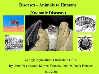
Viral zoonotic diseases Divided into two groups: 1- Arboviruses zoonotic diseases. They are transmitted from animal to human through the bite of arthropode sucking blood vector such as mosquitoes, ticks and sand flies . 2- Non-arboviruses zoonotic diseases. This group include rabies, Marburg, Ebola. Lassa and Hanta viruses
1- Rabies Rabies is also known as hydrophobia. Rabies in Latin means madness . It is a viral zoonotic neurological disease. It is an acute infection of the CNS, that is almost fatal. Wolves, foxes, skunks and raccoons serve as the natural reservoir for the virus . Dogs and cats serve as the most important sources of human infection , because of their close association with humans.
Structure and classification . Family : Rhabdoviridae . genus : lyssa virus The virus is bullet shaped, with helical nucleocapsid. The viral genome is SS-RNA, minus strand. Virion contain the enzyme transcriptase. One major antigenic type exist in nature .
Transmission Rabies is transmitted to human via the bite of rabid animal, mostly dog or cat. Also through contamination of wounds or mucous membranes with infected saliva. The disease does not usually spread from man to man .
Clinical features The incubation period in human depends on the site of the bite( usually between 1 – 4 months ). A bite on the face or neck tend to produce a shorter incubation period than a similar wound on the foot or leg . Clinically the disease in man can be divided into four phases:
Clinical features 1 – the prodromal phase . 2– the excitement phase . 3– the paralytic phase . 4– coma and death .
Clinical features 1--The prodrom may show any of the following: malaise, headache, nausea , vomiting and fever . Usually there is discomfort or paresthesia at the site of the bite . 2--The excitement phase is characterized by: anxiety, agitation, increased nervousness, hyper reactivity, pupillary dilation, increased salivation, painful laryngeal and pharyngeal spasms triggered by swallowing saliva ( hydrophobia ).
Clinical features 3-- Paralytic phase : soon a wide variety of the CNS signs appear including hallucination, lack of coordination, mental confusion and paralysis . 4-- finally coma develop and death . Recovery from rabies is extremely rare. Only six documented cases of human survival from clinical rabies have been reported.
Laboratory diagnosis Rabies diagnosis in human ( during life): by detection of rabies virus –RNA in saliva using reverse transcriptase PCR. In dead animals: by detection of rabies antigens in brain tissue, using direct immun-flourescent technique
Pre-exposure immunization Generally confined to those occupationally at risk such as veterinarians, animal holders, and long term visitors to endemic areas. They should be given 3-doses of the human diploid vaccine one month apart, with a booster dose two years later .Tow booster doses should be given if they are exposed to infection.
Post- exposure, or suspicion of exposure The wound should washed thoroughly with soap and water and alcohol or iodine solution. Patients should be given combined passive and active immunization. Passive immunization: by injecting of human anti-rabies immunoglobulin( RIG ). The dose is 20 Iu/kg, half given around the bite wound and the other half intramuscularly
Post –exposure or suspicion of exposure Active immunization : The main vaccine is the human diploid cell vaccine. Contains: inactivated virus disrupted into subunits. Prepared : in human embryo lung cell. Administered : intramuscularly in 5-doses spaced at 0, 3, 7, 14 & 30 days .
Prevention Stray animals should be destroyed. Vaccination of pet dogs and animals should be mandatory . A live attenuates vaccine is available for immunizing dogs and cats .
2-- Marburg and Ebola virus infections Marburg and Ebola are filoviruses that cause hemorrhagic fever. They cause multiple organ failure with high mortality rate . They are transmitted to human through a direct contact with infected chimpanzees, gorillas and monkeys or their secretions .
Marburg and Ebola virus infection Marburg virus was discovered in 1976 , when outbreaks of hemorrhagic fever occurred simultaneously in Marburg and Frankfurt in Serbia and Germany . Ebola virus was discovered in 1967 and named after the river in Congo. They have been classified in the family filoviridae. They have ss-RNA genomes with negative polarity .
Transmission They infect human and non-human primates (chimpanzees, gorillas and monkeys). Humans are infected when they come in direct contact with infected monkeys and their secretions. Human to human transmission occurs via direct contact with infected blood, secretions and organs of infected person. Health care workers have frequently been infected while attending and caring for patients.
Clinical features The incubation period ranging from 5 – 10 days . The disease is started with fever, myalgia, headache, nausea, vomiting, conjunctival infection. Jaundice and lymphadenopathy. Internal and external bleeding occur within few days. During the second week of infection the patient either begin recovery or develop fatal multiple organ failure. Mortality rate ranges from 25 – 90 % , higher with Ebola.
Laboratory diagnosis Hemorrhagic fever viruses are classified as bio-safety level class four pathogens ( BSL-4 ) . Lab diagnosis must be accomplished under maximum biological containment conditions . The two most commonly used lab methods are, detection of the viral genome in the patient blood using PCR or isolation of the virus in tissue culture followed by identification of the virus .
Treatment and prevention There is no specific anti-viral drug therapy. Treatment is supportive. There is no vaccine available yet for Ebola and Marburg infections.