Download
1 / 41
420 likes | 629 Vues
Ch. 49: Sensory and Motor Mechanism. What is Sensory Reception?. Sensory reception- the detection of the energy of a stimulus by sensory cells Sensory receptors- specialized neurons or epithelial cells that exist singly or in groups with other cell types within sensory organs; eyes and ears

E N D
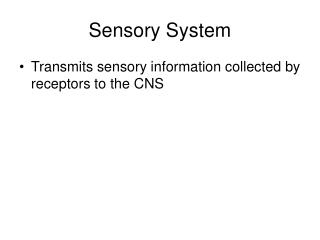
What is Sensory Reception? • Sensory reception- the detection of the energy of a stimulus by sensory cells • Sensory receptors- specialized neurons or epithelial cells that exist singly or in groups with other cell types within sensory organs; eyes and ears • Perception- of the stimuli; interpretation of sensations. Color, smells, sounds and tastes • Sensations- action potentials that reach the brain via sensory neurons • Exteroreceptors- detect stimuli outside the body; heat, light, pressure and chemicals • Interoreceptors- detect stimuli within the body; blood pressure and body position. • Stimuli represent forms of energy, receptor cells convert energy of stimuli into changes in membrane potentials and then transmit signals to the nervous system
Four Functions of Sensory Receptors • Transduction • Amplification • Transmission • Integration
Transduction • Sensory Transduction- detection of a stimulus involves the conversion of stimulus energy into a change in the membrane potential of a receptor cell • Receptor potential- initial response of the sensory receptor to a stimulus is a change in its membrane permeability, resulting in a graded change in membrane potential • Some stimuli such as pressure change stretch the membrane and increase ion flow. • Specific receptor molecules on the membrane of a receptor cell open or close gates to ion channels when the stimulus is present.
Amplification • Amplification- Strengthening of stimulus energy that is otherwise too weak to be carried into the nervous system • Amplification of the signal may occur in accessory structures of a complex sense organ; sound waves are enhanced by a factor of more than 20 before reaching the receptors of the inner ear. But it also may be part of the transduction process. An action potential conducted from the eye to brain has about 100,000 times as much energy as the few photons of light that triggered it, signal transduction pathways in the receptor cells contribute to this amplification
Transmission • Transmission- the conduction of impulses to the central nervous system • The receptor itself is actually a sensory neuron that conducts action potentials to the central nervous system. • Separate receptors- the strength of the stimulus and receptor potential affect the amount of neurotransmitter released by the receptor at synapse with a sensory neuron- determines the frequency of action potentials generated by the sensory neuron which spontaneously generates signals at a low rate, • Stimulus doesn’t actually switch the production of action potentials on or off; it modulates their frequency. The central nervous system is sensitive not only to the presence or absence of a stimulus but also to changes in stimulus intensity
Integration • Integration- processing of information; begins as soon as information is first received. Signals from receptors are integrated through summation of graded potentials, as are those within the nervous system. • Sensory adaptation- a decrease in responsiveness during continued stimulation. Without sensory adaptation, you would feel every beat of your heart and and every bit of clothing on your body. Receptors are selective in the information that they send to the CNS, and adaptations reduces the likelihood that a continuous stimuli will be transmitted.
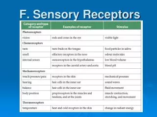
Categorization of Sensory Receptors • Sensory receptors are categorized by the type of energy they transduce • Mechanoreceptors • Pain receptors • Thermoreceptors • Chemoreceptors • Electromagnetic receptors
Muscle Spindle • The mechanoreceptor monitors the length of skeletal muscles. Muscle spindle contains modified muscle fibers attached to sensory neurons and runs parallel to muscle. Muscle is stretched, the fibers of the spindle are also stretched, depolarizing the sensory neurons and triggering action potentials that are transmitted back to the spinal cord.
Hair Cells • Hair cell- common type of mechanoreceptor that detects motion, found in ears of vertebrates and in the lateral line organs of fish and amphibians, where they detect movement relative to their environment. The hairs are specialized cilia or microvilli which project upward from the surface of the hair cell into either an internal compartment; human ear or environment like a pond. When the cilia or microvilli bend in one direction- stretching of the hair cell membrane occurs and it increases its permeability to sodium and potassium ions- increase in the rate of impulse production in a sensory neuron. Cilia bend in opposite direction ion permeability decreases reducing the number of action potentials in the sensory neuron.
Photoreceptors and Vision • Photoreceptors- detect the radiation of visible light, organized into the eyes. • Photoreceptor cells are usually located in a cup. The eye cup is one of the simplest photoreceptor cells.
Compound and Single lens Eyes • Compund eye- insects and crustacians • Ommatidia- several thousand light directors • Single-lens eye- jellies, polychaetes, spiders and mollusks
The Eye • Single Lens Eye: The brain actually sees but the eye is used to take in images that are then sent to the brain to be translated.
The Ear • How does the ear function as a hearing organ? • Converts energy of pressure waves in the surrounding environment into nerve impulses that can be read by the brain.
The Ear: Body Balance and Equilibrium • How does the functions of the ear maintain body balance and equilibrium? • The utricle and saccule in the vestibule and the semicircular canals contain hair cells sensetive to balance and body position.
Human endoskeleton 206bones
The skeleton • Support • Protection • Movement
Endoskeleton • Endoskeleton- hard supporting elements, like bones, buried within the soft tissues of an animal.
Exoskeletons • Exoskeleton- hard encasement deposited on the surface of an animal.
Coelom • Coelom fluid acts as a hydrostatic skeleton • Using muscles an earthworm can change the shape of the segments that divide the coelmic cavity
Arthropod Exoskeleton • Cuticle- nonliving coat secreated by the epidermis. Muscles are attached to cuticle. Cuticle consists of chitin and chitin fibrils are embedded in a protein matrix forming a composite material both strong and flexible. Cuticle hardened by organic compounds- cross-link proteins of exoskeleton.
The Trigger: motor neurons • Motor neuron triggers muscle contraction
Muscle • Skeletal- attached to the bones and is responsible for their movement, characterized by a hierarchy of smaller and smaller parallel units. • Cardiac Muscle- muscle that is found only in the heart • Smooth Muscle- lacking the striations of skeletal and cardiac muscle because of the uniform distribution of myosin filaments
Muscle Arrangement • Muscles do work by contracting • skeletal muscles come in antagonistic pairs • flexor vs. extensor • contracting = shortening • move skeletal parts • tendons • connect bone to muscle • ligaments • connect bone to bone
Muscle Contractions • The nervous system controls the strength of muscle contractions by determining how many motor units are activated at a given time and whether small or large motor units are activated.
Sarcoplasmic reticulum • Sarcoplasm • muscle cell cytoplasm • contains many mitochondria • Sarcoplasmic reticulum (SR) • organelle similar to ER • network of tubes • stores Ca+2 • Ca+2 released from SR through channels • Ca+2 pumps then restore Ca+2 to SR • remove Ca+2 from cytosol • pumps use ATP Ca+2 ATPase of SR
Sacromere • In a relaxed Sacromere the thin and thick filaments barely overlap while in a contracted sacromere the thick and thin filaments do overlap.
Striated skeletal muscle A band = thick filaments = myosin I band = thin filaments = actin Z Z
Muscle filaments & Sarcomere • Interacting proteins • thin filaments • braided strands of actin & tropomyosin coiled together • thick filaments • myosin molecules
Thin and Thick Filaments Myosin tails together & heads pointed away from center of sarcomere Thick Filament Thin Filament
Interaction of thick & thin filaments • Cross bridges formed between myosin heads (thick filaments) & actin (thin flaments) cause the muscle to shorten (contract)
Cross bridge cycle • Cleaving ATP allows myosin head to bind to actin filament
Sliding Filament Model • Filament do not change length in muscle contraction • Filament overlap each other • Reducing the I band and the H band
Calcium and Muscle Contractions • Calcium bound with troponin it causes the myosin binding sites on actin to be exposed • Calcium allows thick and thin filaments to slide and overlap • Sacroplasmic reteculum controls the calcium concentration
Skeletal Muscle Contractions • 1. Ach released from neuron diffuses along the synaptic cleft and attaches to Ach receptors on the muscle cells plasma membrane • 2. Action potential is propagrated along the plasma membrane and down the T tubules • 3. Action potential triggers Ca2+ released from the SR. • 4. Calcium Ions Bind to troponin; troponin changes shape, actin active sites exposed
Skeletal Muscle Contraction Cont. • 5. Myosin cross-bridges alternately attach to actin and detach, pulling actin filaments towards the center of the sarcomere. ATP powers the sliding of the filaments • 6. Cytosolic Ca2+ is removed by active transport into the SR after the action potential ends. • 7. Tropomyosin blockage is restored, covering actin active site. The contraction ends and the muscle fiber relaxes
How it all works… • Action potential causes Ca+2 release from SR • Ca+2 binds to troponin • Troponin moves tropomyosin • Tropomyosin uncovers myosin binding site on actin • Myosin binds actin • uses ATP to "rachet" once • releases, "unratchets" & binds to next actin • Myosin pulls actin chain along • Sarcomere shortens • Z discs move closer together • Whole fiber shortens contraction! • Ca+2 pumps restore Ca+2 to SR relaxation! • pumps use ATP
Fast twitch & slow twitch muscles • Slow twitch muscle fibers • contract slowly, but keep going for a long time • more mitochondria for aerobic respiration • less SR Ca+2 remains in cytosol longer • long distance runner • “dark” meat = more blood vessels • Fast twitch muscle fibers • contract quickly, but get tired rapidly • store more glycogen for anaerobic respiration • sprinter • “white” meat