Download
1 / 13
130 likes | 225 Vues
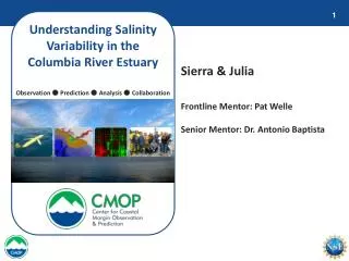
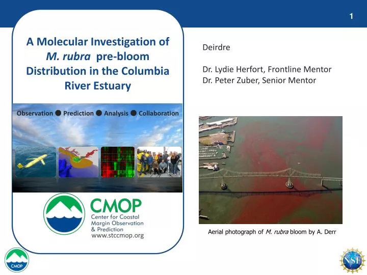
A Molecular Investigation of M. rubra pre-bloom Distribution in the Columbia River Estuary. Deirdre Dr. Lydie Herfort, Frontline Mentor Dr. Peter Zuber, Senior Mentor. Observation ● Prediction ● Analysis ● Collaboration. Aerial photograph of M. rubra bloom by A. Derr.

E N D
A Molecular Investigation of M. rubra pre-bloom Distribution in the Columbia River Estuary Deirdre Dr. Lydie Herfort, Frontline Mentor Dr. Peter Zuber, Senior Mentor Observation ● Prediction ● Analysis ● Collaboration Aerial photograph of M. rubra bloom by A. Derr www.stccmop.org
What is Myrionecta rubra? • Mixotrophic ciliate, most likely of marine origin • Forms non-toxic red tides in estuaries, fjords, and other coastal margin environments • Photosynthesizes through use of acquired chloroplasts of cryptophyte prey • Karoklepty (predation) • Symbiotic co-evolution Microscope image of cryptophyte prey (T. Peterson) M. rubra under transmitted light (left) & epoflourescence microscopy (right) by D. Stoecker, University of Maryland
M. rubra blooms in the Columbia River estuary • Blooms from late July to October • Based on ‘18S-28S’ rRNA gene analysis, a single variant leads to blooms each year (variant B) • Only one cryptophyte, Teleaulax amphioxea, is associated with M. rubra variant B Aerial photography of M. rubra bloom by A. Derr, 2008
M. rubra in Oceanic Waters • At least five different variants detected in coastal waters based on ‘18S-28S’ rRNA gene analysis Sites of sequence polymorphisms of M. rubra partial ‘18S-28S’ rRNA gene sequences
M. rubra in Oceanic Waters, continued • Water samples collected during CMOP May-June cruise 2010 • FlowCAM analysis of 15 mL of water showed M. rubra in only two locations FlowCAM images of M. rubra among phytoplankton assemblages (T. Peterson)
Question & Research Goal Question: • FlowCAM – 15 mL of sample water • Molecular analysis – 1-4 L of sample water • Is molecular analysis a more sensitive approach? Goal: • Determine M. rubra presence in coastal water samples using molecular identification methods • Polymerase Chain Reaction (PCR) DNA amplification with M. rubra specific primers • Agarose gel electrophoresis to visualize PCR products
Data Collection Methods • Analyze 18 samples taken from the CMOP May-June cruise 2010 at varying depths and locations • Extract nucleic acid from filtered water samples using a phenol/chloroform extraction method • Use 18S rRNA gene PCR primers (EukA & EukB) for general identification of microbial eukaryotes • Use ‘18S-28S’ rRNA gene PCR primers (MR18Sf & MR28Sr) specific to M. rubra • Amplifies M. rubra Internal Transcribed Spacer gene region (below) • Run all PCR products on agarose electrophoresis gel to visualize PCR products
Results • Nucleic acid successfully extracted from filtered water samples • Microbial eukaryotes detected in 14 / 18 samples • M. rubra detected in 17 / 18 samples, even when not detected by FlowCAM
Results, continued • Key • Gave a PCR signal • Gave no PCR signal • (Surface – Left • Middle – Center • Bottom – Right) Eukaryotes M. rubra
Conclusions • M. rubra present in most coastal samples during pre-bloom season • M. rubra not detected by FlowCAM analysis because it is likely present in low abundance Preparing PCR products for an agarose electrophoresis gel (J. Schilling)
Sampling Experience • Went water sampling in Astoria and Ilwaco Harbor with Sheedra Futrell and Dr. Lydie Herfort
Future Work • Continue monitoring M. rubra presence during non-blooming periods (Nov. – Jun.) • Identify which variant of M. rubra is most common in oceanic samples • Develop an alternative method to gene sequencing for identification of M. rubra variants • Culture the five known variants of M. rubra to use as positives for PCRs
Thank You! • My frontline mentor, Dr. Lydie Herfort • My senior mentor, Dr. Peter Zuber • Vikki Campbell • Karen Wegner • Dr. Antonio Baptista