Download
1 / 46
510 likes | 967 Vues
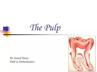
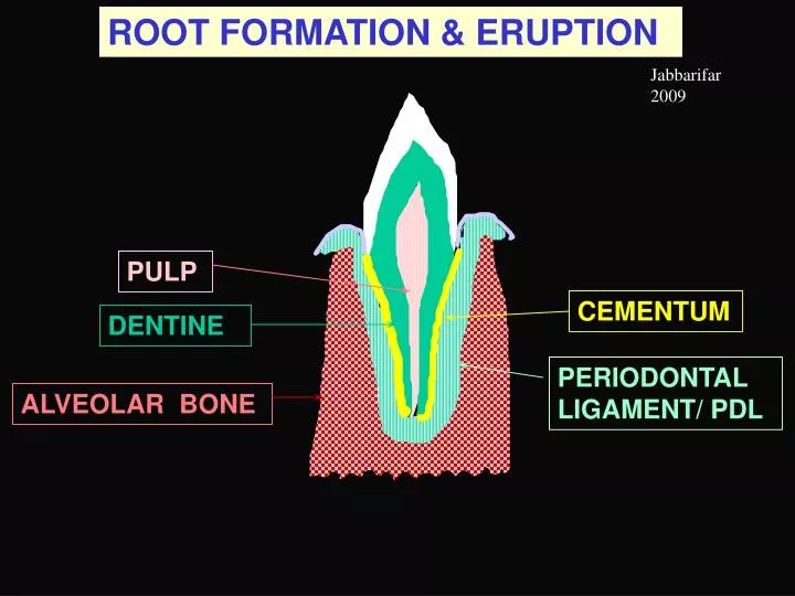
ROOT FORMATION & ERUPTION. Jabbarifar 2009. PULP. CEMENTUM. DENTINE. PERIODONTAL LIGAMENT/ PDL. ALVEOLAR BONE. What has to be controlled. Number of roots. Shapes of root. Length of root. Four tissues in sequence. Pulp Dentine Cementum Ligament. Organize surroundings.

E N D
ROOT FORMATION & ERUPTION Jabbarifar 2009 PULP CEMENTUM DENTINE PERIODONTAL LIGAMENT/ PDL ALVEOLAR BONE
What has to be controlled Number of roots Shapes of root Length of root Four tissues in sequence Pulp Dentine Cementum Ligament Organize surroundings Fasten tooth to surroundings Times of eruption & coordinated Shedding of teeth e.g., cementum with PDL with bone Successional teeth
TOOTH TISSUES: Cell Sources DENTAL LAMINA DENTAL ORGAN DENTAL PAPILLA DENTAL SAC/FOLLICLE ALVEOLAR BONE TOOTH ENAMEL Ameloblasts DENTINE Odontoblasts PULP CT cells CEMENTUM Cementoblasts PDL Fibroblasts A BONE Osteoblasts & ‘clasts Crest
MECHANISMS OF ERUPTION Construction & Reorganization of PDL Formation of the root Deposition of alveolar bone? Remodelling of bone overall FURTHER INFLUENCES from: tooth/teeth in occlusion; muscle actions
TOOTH GERM: next steps Outer dental epithelium approaches inner DENTAL LAMINA upper part degenerates lower forms 2nd bud Stellate reticulum reduces over cusp Knot cells signal to papilla Stratum intermedium DENTAL PAPILLA Inner & outer dental epithelia join to form cervical loop DENTAL SAC/FOLLICLE
LATE CROWN FORMATION cusp enamelformed byameloblasts First reduction in enamel epithelium:active ameloblasts& compacted outer epithelium, stellate-reticulum cells & stratum intermedium Dentine DENTAL PAPILLA become pulp remaining Stellate reticulum Cervical loop: inner & outer epithelium DENTAL SAC still quiescent
END OF CROWN FORMATION Ameloblasts willfinish full thickness of cusp enamel & reduce in height Dentine widens DENTAL PAPILLA become pulp process proceeds downs Stellate reticulum follows Cervical loop down then stops: Crown defined Odontoblast recruitment site Cervical loop:
ROOT FORMATION ENAMEL REDUCED DENTAL EPITHELIUM CROWN DENTINE Where Stellate reticulum stopped, the cervical loop continued to grow down, but as PULP HERTWIG’S ROOT SHEATH & its ROOT Odontoblast recruitment site Epithelial diaphragm
FURTHER ROOT FORMATION REDUCED DENTAL EPITHELIUM Root sheath breaks up, allowing sac mesenchymalcells to contact root dentine ENAMEL DENTINE PULP Other sac mesenchymalcells construct PDL & some alveolar bone HERTWIG’S ROOT SHEATH grows to lengthen root Fibroblasts Odontoblast recruitment site Epithelial diaphragm
ROOT FORMATION: Multirooted ROOT SHEATH & ITS DIAPHRAGM widens & constricts to create two diaphragms to define two roots CROWN ENAMEL DENTINE Cross-sections PULP ROOT ROOT SHEATH Epithelial diaphragm
ROOT FORMATION: Multirooted CROWN ENAMEL DENTINE PULP ROOT ROOT SHEATH Epithelial diaphragm Thus, one dental organ can produce two or three roots Similarly, one dental organ can produce two or more cusps, using multiple enamel knots
REITERATIVE SIGNALING V REDUCED DENTAL EPITHELIUM ENAMEL DENTINE Root sheath breaks up & lifts, allowing sac mesenchymalcells to contact root dentine Dentine &/or Epithelial root sheathinduces mesenchymal cellsto become cementoblasts PULP Odontoblast recruitment site byroot sheath: pulp signaling
JAW & TOOTH DEVELOPMENT early arch BONE starting BUCCAL PLATE DENTAL LAMINA BONE starting LINGUAL PLATE 10 TOOTH GERM 20 Successional TOOTH GERM WALLS OF BONY TROUGH OF DEVELOPING MANDIBLE SYMPHYSEAL CARTILAGE
JAW & TOOTH DEVELOPMENT processes DENTAL LAMINA will grow back to form germs for 3 permanent molars (5th e m) BONE starting BUCCAL PLATE grows up more than lingual BONE starting LINGUAL PLATE 10 TOOTH GERM 20 Successional TOOTH GERM on lingual side of 10 Interradicular septum grows between roots of multirooted teeth Bony wall grows around & encloses 20 TOOTH GERM in a crypt SYMPHYSEAL CARTILAGE will be replaced by bone Interdental septum grows across trough to separate teeth
TOOTH & MANDIBLE DEVELOPMENT Oral ectoderm TONGUE DENTAL SAC 10 TOOTH ALVEOLAR BONE 20 TOOTH GERM MECKEL’S CARTILAGE ALVEOLAR NERVE
MANDIBLE DEVELOPMENT DENTAL SAC contributes also to alveolar bone Remodeling will bring erupting 1o tooth over developing 2o Alveolar crest grows up Bony plate grows up to enclose 2nd tooth germ in a CRYPT 10 TOOTH Bone added to base of alveolus for tooth eruption 20 TOOTH GERM Bone grows over alveolar nerve & vessels MECKEL’S CARTILAGE regresses & not used to form mandible Alveolus becomes distinct from BODY
TOOTH & MANDIBLE DEVELOPMENT - Next Reduced enamel epithelium fused with gingiva TONGUE DENTAL SAC Higher alveolar bone - i.e. deeper socket 10 TOOTH ALVEOLAR BONE Longer root with cementum forming 20 TOOTH GERM More advanced 2nd tooth ALVEOLAR NERVE MECKEL’S CARTILAGE Denser alveolar bone & more body-alveolus distinction Remodeling brings erupting 1o tooth over developing 2o Meckel’s cartilage gone
TOOTH EMERGENCE CUTICLE will wear away GINGIVAL EPITHELIUM still fusing with REDUCED DENTAL EPITHELIUM ENAMEL
ORGANIC ENAMEL SURFACES CUTICLE will wear away PELLICLE of glycoproteins etc is acquired later from saliva PLAQUE the biofilm of many kinds of bacteria then attaches to the pellicle, & later mineralizes - tartar
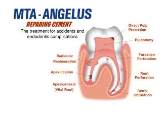
LATE ERUPTING TOOTH Epithelial diaphragm ROOT SHEATH GINGIVA CEMENTUM BONE PDL DENTINE ENAMEL PULP Rests of Mallassez remnants of Root sheath Cementum starting as sheath breaks down
LATE ERUPTING TOOTH ENAMEL Rests of Mallassez remnants of Root sheath DENTINE GINGIVA PULP CEMENTUM PDL ROOT SHEATH Epithelial diaphragm Cementum starting as sheath breaks down BONE
MANDIBULAR CENTRAL INCISORS at 2 y Deciduous tooth Gingiva PDL Permanent tooth ALVEOLAR BONE BODY of MANDIBLE Cortical plate dense bone
Go Gubba Gubernacular cord of fibrous tissue Gubernacular cord runs through a canal left in the bony crypt, where the dental lamina extended down to establish the germ for the 2nd tooth Permanent tooth
Deciduous tooth MANDIBULAR CENTRAL INCISORS at 2 y - Bone Resorption of bone & deciduous root will start here Permanent tooth Spongy/ cancellous bone Cortical plate dense bone
Close to EXFOLIATION of Deciduous/10 Tooth 10/Deciduous tooth Odontoclasts have resorbed most of deciduous root PDL attachment is surprising persistent Pulp is left alive Bone remodellling has brought 20 tooth under 10 20 tooth Bone trabeculae added by layers at base of alveolus 20 tooth would be LARGER than shown
LATE ERUPTING TOOTH: Origins Epithelial diaphragm ROOT SHEATH Dental organ GINGIVA Oral Ectoderm CEMENTUM Dental sac PDL DENTINE ENAMEL PULP Rests of Mallassez remnants of Root sheath BONE BONE Arch Mesenchyme & Dental sac PDL Dental sac
WHY STILL ERUPTING APEX INCOMPLETE BONE PDL DENTINE ENAMEL PULP Cementum not to apex Epithelial diaphragm present Pulp chamber wide (no apical taper) Immature connective tissue & Bone forming in base of alveolus
STARTING EXFOLIATION of DECIDUOUS MOLAR I ENAMEL DENTINE ALVEOLAR BONE PULP PDL Root resorption by osteoclasts Inter-radicular septum of bone also houses 2nd tooth germ & is its crypt Permanent Tooth under deciduous molar, & between its roots
EXFOLIATION of DECIDUOUS MOLAR II Crypt bone eroded here ENAMEL DENTINE ALVEOLAR BONE PDL Resorbed dentine partly repaired by new cementum Focal erosion along this line leaves a ROOT FRAGMENT which may be retained PDL is disrupted in regions of root resorption & repair
EXFOLIATION of DECIDUOUS MOLAR III ENAMEL DENTINE Erosion of bone and the deciduous root is not steady & continuous, but may cease briefly, when some repair of eroded cementum & dentine can occur (by cementum). Bone remodelling also goes on, and the alveolus and crypt are changing all the time - repeated all along the jaw
Occlusal wear FUNCTIONAL ERUPTION & TOOTH MOVEMENT Osteoclasts resorbing bone Osteoblasts laying down bundle bone Bony interdental septum PDL fibers incorporated in bone as Sharpey’s fibers Cellular cementum added to apex Compensates for occlusal wear? Basil
Tooth drifts mesially by combined actions of osteoclasts & osteoblasts moving bone, taking tooth with it TOOTH MOVEMENT Osteoblasts laying down bundle bone Osteoclasts resorbing bone Basil
TOOTH MOVEMENT Tooth drifts mesially by combined actions ofosteoclasts&osteoblastsmovingbone, taking tooth with it Earlier bone position Basil
TOOTH ERUPTION Pre-oral phase Intra-oral phase
TOOTH ERUPTION Once the teeth meet inocclusion, their further eruption separates the jaws Once the teeth meet in occlusion, they influence each other mechanically
PERIODONTITIS TOOTH EPITHELIAL ATTACHMENT- unstable, loosens & migrates down, & allows bacteria into CONNECTIVE TISSUE resulting in chronic infection & inflammation & systemic spread of bacteria & loss of teeth GINGIVA Periodontal ligament Alveolar bone
PASSIVE ERUPTION Gingival recession onto & down the cementum with loss of alveolar-crest bone Raising the banana, then peeling the banana
Fate of exposed cementum & dentinal consequences & reactions PULP Cementum readily abraded & eaten by oral acids DEAD TRACT in Dentine - wide, empty dentinal tubules easily colonized by bacteria REPARATIVE DENTINE - response to caries/erosion
TOOTH MOVEMENTS Occurring in eruption & use TILTING By root growth & bone remodelling AXIAL - in long axis of the tooth DRIFTING e.g., mesially, laterally By bone remodelling & PDL reorganization ROTATORY Combinations of these four movements frequently occur Basil
TOOTH MOVEMENT 2 TILTING Tooth tilts by combined actions of both osteoclasts&osteoblasts on bone of each side of socket Earlier bone position Basil
3rd MOLAR’S TILTING ERUPTION 2nd 2nd 3rd 3rd TILTING mechanism may be useful, e.g., in bringing upright the third molar that starts tilted Failure can lead to an impacted molar still within the bone
YOUNG CHILD’S ERUPTION SEQUENCE 10 2nd Molar YEARS 0 1 2 3 4 5 6 7 KEY Time of emergence Crown forming Root forming Deciduous Permanent 20 Incisor 20 Cuspid 20 2nd PreMolar
YOUNG CHILD’S ERUPTION SEQUENCE 10 2nd Molar YEARS 0 1 2 3 4 5 6 7 10 Incisor 10 Cuspid 20 Incisor 20 Cuspid 20 2nd PreMolar
5-yr CHILD’S DENTITION: 0ne arch Oral Pre-oral 5 deciduous teeth working, but 1o incisor root is being resorbed 7 successional/succedaneous teeth developing pre-orally dental lamina for 3rd molar
5-yr CHILD’S DENTITION: 0ne arch Oral Pre-oral These 12 “teeth” require a very coordinated remodelling of the bone (& PDL) supporting & enclosing them
ERUPTION: Problems Delayed eruption Early eruption Missing tooth Impaction - failure to erupt e.g., from too little gap after premature loss of deciduous tooth Retained root fragment Malocclusion Infra-occlusion (not high enough) Excessive drift Tilting (can occur early from germ rotation)