Download
1 / 30
420 likes | 1.03k Vues
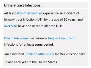
Urinary Tract Tumors. 2%-3% of all cancers in adults . The most common malignant tumor of the kidney is renal cell carcinoma . followed in frequency by nephroblastoma ( Wilms tumor) and by primary tumors of the calyces and pelvis.

E N D
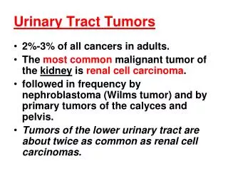
Urinary Tract Tumors • 2%-3% of all cancers in adults. • The most common malignant tumor of the kidney is renal cell carcinoma. • followed in frequency by nephroblastoma (Wilms tumor) and by primary tumors of the calyces and pelvis. • Tumors of the lower urinary tract are about twiceas common as renal cell carcinomas.
Renal Cell Carcinoma (RCC) • are derived from the renal tubular epithelium. • located predominantly in the cortex. • 2%-3% of all cancers in adults. • 80%-85% of all primary malignant tumors of the kidney. • 30,000 cases per year. • 40% of patients die of the disease. • 6th-7th decades. • M:F 2:1
Predisposing factors • 1- smoking • 2- hypertension • 3- obesity • 4- occupational exposure to cadmium. • Smokers who are exposed to cadmium have a particularly high incidence of renal cell carcinomas. • 5- chronic dialysis & acquired polycystic disease -The risk of developing renal cell cancer is increased 30-fold
New classification based on the molecular origins of these tumors • 1-Clear Cell Carcinomas • 2-Papillary Renal Cell Carcinomas • 3-Chromophobe Renal Carcinomas
1-Clear Cell Carcinomas • are the most common type (70%- 80% of RCC). • Histologically, they are made up of cells with clear or granular cytoplasm. • Forms of clear cell renal carcinoma: • 1-Sporadic • 2-Familial • 3-in association with von Hippel-Lindau (VHL) disease
VHL disease • VHL gene is tumor suppressor gene involved in limiting the angiogenic response to hypoxia; thus, its absence may lead to increased angiogenesis and tumor growth • is autosomal dominant and is characterized by predisposition to a variety of neoplasms: • 1- hemangioblastomas of the cerebellum and retinal angiomas. • 2- bilateral renal cysts • 3- bilateral & multiple clear cell carcinomas (40%-60% of individuals). • 4- Pheochromocytoma
Those with VHL syndrome inherit a germ-line mutation of the VHL gene on chromosome 3p25 and lose the second allele by somatic mutation. • The VHL gene is also involved in the majority of sporadic clear cell carcinomas (60%). • homozygous loss of the VHL gene seems to be the common underlying molecular abnormality in both sporadic and familial forms of clear cell carcinomas
2-Papillary Renal Cell Carcinomas • 10% to 15% of all renal cancers. • show a papillary growth pattern. • are frequently multifocal and bilateral and appear as early-stage tumors. • familial and sporadic forms. • papillary renal cancers have no abnormality of chromosome 3. • The gene involved in papillary renal cell cancers is the MET proto-oncogene, located on chromosome 7q31. • The MET gene is a tyrosine kinase receptor for the growth factor called hepatocyte growth factor (HGF). • increased gene dosage of the MET gene due to duplications of chromosome 7 seems to spur abnormal growth in the proximal tubular epithelial cell precursors of papillary carcinomas.
familial cases: • trisomyof chromosome 7 • activating mutations of the MET gene. • sporadic cases: • duplication or trisomy of chromosome 7 • but there is no mutation of the MET gene. • chromosomal translocation involving chromosome 8q24 close to the c-MYC gene, is also associated with some cases of papillary carcinoma
3-Chromophobe Renal Carcinomas • the least common (5% of all RCC). • They arise from intercalated cells of collecting ducts. • the tumor cells stain more darkly (i.e., they are less clear) than cells in clear cell carcinomas. • These tumors are unique in having multiple losses of entire chromosomes, including chromosomes 1, 2, 6, 10, 13, 17, and 21. • they show extreme hypodiploidy. • Because of multiple losses, the "critical hit" has not been determined. • chromophobe renal cancers have a good prognosis.
Renal cell carcinoma: typical cross-section of yellowish, spherical neoplasm in one pole of the kidney. Note the tumor in the dilated, thrombosed renal vein.
Renal cell carcinomaHigh-power detail of the clear cell pattern
Clinical Course 1-the most frequent presenting manifestation is hematuria( in more than 50% of cases). • Macroscopic hematuria tends to be intermittent and fleeting superimposed on a steady microscopic hematuria. 2-Less commonly the tumor may present flank pain and a palpable mass. The characteristictriad of : • painless hematuria • a palpable abdominal mass • dull flank pain 3-Fever 4-Polycythemia ( 5% to 10% of cases): It results from elaboration of erythropoietin by the renal tumor.
Paraneoplasticsyndromes: • 1-hypercalcemia • 2-Hypertension • 3-Cushing syndrome • 4-feminization or masculinization • The prevalent locations for metastases are the lungs and the bones. • may invade the renal vein and grow within this vessel, sometimes extending as far as the inferior vena cava and even into the right side of the heart.
Wilms Tumor • it is the 3rd most common solid organ cancer in children < age of 10 years. • contain cells and tissue components all derived from the mesoderm. • may arise sporadically or familial (susceptibility to tumorigenesis inherited as an autosomal dominant trait). • Mutations involve WT1 gene. • The tumor shows attempts to form primitive glomerular and tubular structures
Wilm's tumor nests and sheets of dark blue cells at the left with compressed normal renal parenchyma at the right.
The tumor shows attempts to form primitive glomerular and tubular structures
Bladder cancer • accounts for 7% of cancers and 3% of cancer deaths in the USA. - (90%) are urothelial carcinomas. - squamous cell carcinomas (SCC): 3% to 7% - Adenocarcinoma: rare
Urothelial tumors • Tumors in the collecting system above the bladder are relatively uncommon. • These tumors are classified into : • 1 benign papilloma. • 2-papillary urothelialneoplasms of low grade • 3-papillary urothelial carcinoma of high grade
Urothelial (transitional) cell carcinomas • range from papillary to flat, noninvasive to invasive, and low grade to high grade. • Low-grade carcinomas are always papillary and are rarely invasive. • they may recur after removal. • Increasing degrees of cellular atypia and anaplasia • High-grade cancers can be papillary or occasionally flat.
squamous cell carcinomas • only 5% of bladder cancers are squamous cell carcinomas. • in situ stage of bladder carcinoma can be recognized • wide areas of atypical hyperplasia and dysplasia may be present. • Associated with chronic inflammation and stone formation, also Schistosomiasis infection
Clinical Course • Painless hematuria is the dominant clinical presentation of all tumors. • M:F 3:1 • 50 to70 years. • A wide variety of genetic abnormalities are seen in bladder cancers: • chromosome 9 mutations in several genes on (including p16) • p53 • FGFR3
Predisposing factors : • Bladder cancer is not familial. • factors implicated in the causation: • 1- 50 X more common in those exposed to β-naphthylamine. • 2-Cigarette smoking. • 3-Chronic cystitis. • 4-Schistosomiasis of the bladder. • 5-drugs as cyclophosphamide.
The clinical significance of bladder tumors depends on : • 1- histologic grade. • 2-differentiation . • 3-the depth of invasion of the lesion. • all tend to recur after removal except for benign papillomas. • Lesions that invade the ureteral or urethral orifices cause urinary tract obstruction. • Prognosis • low-grade shallow lesions have good prognosis. • with deep penetration of the bladder wall the 5-year survival rate is less than 20%. • Overall 5-year survival is 57%.
The extent of invasion and spread (staging) at the time of initial diagnosis is the most important prognostic factor • Treatment: • transurethral resection is both diagnostic and therapeutic • bacilleCalmette-Guérin (BCG)granulomatousreaction;triggers local immune response • Follow-up for recurrence with periodic cystoscopy and urine cytologic studies for the rest of their lives. • Radical cystectomy and chemotherapy for advanced cases
Papillary urothelial carcinoma of ureter & renal pelvis • 5% to 10% of primary renal tumors. • Painless hematuria is the most characteristic feature of these lesions. • Depending on critical location they produce pain in the costovertebral angle as hydronephrosis develops. • Infiltration of the walls of the pelvis, calyces, and renal vein worsens the prognosis. • Despite removal of the tumor by nephrectomy, fewer than 50% of patients survive for 5 years. • Cancer of the ureter is fortunately the rarest of the tumors of the collecting system. • The 5-year survival rate is less than 10%.
The cut surfaces of the kidney demonstrate normal cortex and medulla, but the calyces show focal papillary tumor masses of transitional cell carcinoma.