Download
1 / 37
390 likes | 623 Vues
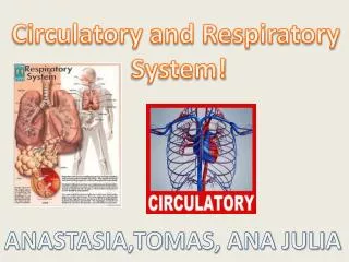
Chapter 37 Circulatory System Respiratory System. 37.1 Circulatory System A. Transportation system of living organisms : Oxygen from the lungs to the cells of the body Nitrogenous wastes to the kidneys for removal Carbon dioxide waste from cells to the lungs for removal

E N D
37.1 Circulatory System A. Transportation system of living organisms: • Oxygen from the lungs to the cells of the body • Nitrogenous wastes to the kidneys for removal • Carbon dioxide waste from cells to the lungs for removal • Food nutrients from the intestines to cells for energy
B. Circulatory system is composed of: 1. Heart • Cardiac muscle which is enclosed in protective tissue called pericardium • Contractions pump blood through the C. S. average 72 times/min • The heart has its own blood supply through the coronary arteries bark scorpion venom • Heart is divided in the middle by the septum
4 heart chambers • Atria: 2 upper chambers • that receive blood into • the heart. Receiving chambers • Ventricle: 2 lower chambers that • pump blood out of the • heart. Pumping chambers. • Ventricles have thick muscle to • pump to the lungs/body septum
heart animations • heart parts • There are valves between the atria and ventricles • tricuspid & mitral valves separate the atria • from ventricles. Prevent back flow into 2 atria. • pulmonary & aortic valves separate the major vessels leaving the heart from the 2 ventricles Good flow through heart
septum Inferior vena cava
Circulation Heart functions as 2 separate pumps that circulate blood in 2 circuits around the body a. Pulmonary Circulation: heart lungs heart - Right ventricle pumps blood to the lungs - high CO2 - blood enters the lungs and absorbs oxygen and releases carbon dioxide. - this oxygenated blood then returns to the left side of the heart
b. Systemic Circulation hearr body heart - The left ventricle pumps blood to the whole body delivering oxygen to the cells. - Flow goes back to the right side of the heart with CO2 rich blood Circulation PH
Circulation Right side Left side
Pacemakers: 2 bundles of nerves in the heart that when stimulated, causes the atria and ventricles to contract. SA node: contracts the atria to force blood into the ventricles AV node: contracts Ventricles top force blood out of the heart
2. Vessels: blood flows through three types of vessels Arteries - Carry blood AWAY from the heart. (oxygenated except pulmonary art) - Aorta is thick to withstand pressure of bl. flow Capillaries - Bring nutrients and oxygen to tissues of organs and absorb carbon dioxide and waste from tissues and organs. - One cell layer thick to allow diffusion Veins - Carry blood TO heart. (deoxygenated except pulmonary vein from lung) - Have valves to keep blood flowing up toward the heart.
Blood Vessels Oxygen flows from the heart through arteries and capillaries into tissues and carbon dioxide flows into the capillaries and to the veins back to the heart angioplasty
Blood Pressure: - Force of blood on arteries - Systolic number in blood pressure is force on arteries when ventricles contract. - Diastolic number is force on arteries when ventricles are relaxed. - Normal – 120/80 - blood pressure
E. Blood – a. Plasma: yellow liquid – blood clotting, proteins b. Red blood cells: transport oxygen. Contain hemoglobin. Iron protein that binds o2 c. White blood cells: fight infection d. Platelets: blood clotting proteins c
plasma Red blood cells White BC and Platelets Lymphatic system
Types of White Blood Cells Section 37-2 Cell Type Neutrophils Eosinophils Basophils Monocytes Lymphocytes Function Engulf and destroy small bacteria and foreign substances Attack parasites; limit inflammation associated with allergic reactions Release histamines that cause inflammation; release anticoagulants, which prevent blood clots Give rise to leukocytes that engulf and destroy large bacteria and substances Some destroy foreign cells by causing their membranes to rupture; some develop into cells that produce antibodies, which target specific foreign substances
37.3 Respiratory System A. Respiration exchanges gas between organisms their environment. (cellular respiration is converting sugars to ATP in the mito.) B. Air follows the following pathway: pharynx: Air moves through the nose to a tube at the back of the mouth. passage of both air & food. Trachea: At the top of the trachea is a flap of tissue called the epiglottis. It covers trachea when eating so that food goes down the esophagus and not the trachea. -
cilia & mucus along the nose and pharynx trap dust and smoke. Cilia pushes junk up. where it is either spit out or swallowed. Larynx: voice box – 2 folds of tissue that form a slit. When air moves through them, sound is made. Bronchi: 2 large passages lead air to each lung and then to smaller tubes called bronchioles. Surrounded by smooth muscle.
bronchioles - smaller branched passageways that lead from each bronchus to clusters of alveoli. - they help increase surface area of alveoli in the lungs
Alveoli: Small sacs 150 million/lung richly supplied with capillaries actual site of gas exchange huge surface area (60 square meters) Clustered at end of bronchioles alveoli
Figure 37-13 The Respiratory System Section 37-3
Alveoli/capillaries - Gas Exchange higher conc of O2 in inhaled air than exhaled air and higher conc CO2 in exhaled air than inhaled air. O2 diffuses from alveoli into capillaries CO2 diffuses from capillaries to the alveoli respiration PH respiration O2 CO2 Capillary
Red blood cells moving through capillaries around the alveoli of the lung.
C. Breathing diaphram & breathing • No muscles connected to the lungs, moving them. • Lungs expand due to difference in pressure between the chest cavity and the atmosphere. • Diaphragm: - when you breath in, the diaphragm contracts and the rib cage expands. - the volume of the chest cavity (between the lung and rib cage) increases - because the cavity is sealed – a vacuum is formed and the chest cavity pressure decreases - atmospheric pressure is greater, so air is forced into the lungs
As the diaphragm relaxes, the pressure increases in the chest cavity and air is forced out. Air exhaled Air inhaled Rib cage lowers Rib cage rises Diaphragm Diaphragm Exhalation Inhalation
D. Control of breathing • Breathing can be voluntary • Much of breathing is autonomic. • The medulla oblongata (brain stem) monitors the levels of CO2 in the blood. • If CO2 levels increase, impulses to the diaphragm cause more contractions and more air is brought in. A nerve travels from the brain to the diaph. • The higher the CO2 level, the stronger the impulse. • When you exercise, you actually breath faster to release CO2, not so much to get O2.
Respiratory Diseases • Bronchitis: inflamation if bronchi • Emphysema: loss of elasticity of lung tissue alveoli can’t expand for gas ex. tobacco damages the tissue
Asthma: narrowing of the bronchi and bronchioles due to the constriction of muscles around the airways. Environmental, genetic? • Cystic fibrosis: recessive, autosomal genetic disease in which lungs collect mucous and cause multiple infections.