Download
1 / 15
200 likes | 613 Vues
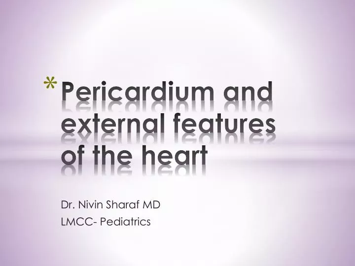
Pericardium and external features of the heart. Dr. Nivin Sharaf MD LMCC- Pediatrics. By the end of this lectures the students should be able to: Discuss the gross anatomy of the pericardium Identify pericardial sac Differentiate between terms: serous pericardium, fibrous pericardium

E N D
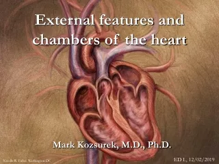
Pericardium and external features of the heart Dr. Nivin Sharaf MD LMCC- Pediatrics
By the end of this lectures the students should be able to: • Discuss the gross anatomy of the pericardium • Identify pericardial sac • Differentiate between terms: serous pericardium, fibrous pericardium • Recognize the importance of pericardial sinuses Objectives
Pericardium is a fibro serous sac that encloses the heart and the roots of the great vessels • Extends between 2nd- 6th costal cartilages • Anterior to 5th to 8th Thoracic vertebrae Definition
Strong fibrous part of the sac • Firmly attached below to the central tendon of diaphragm • Attached to the back of the sternum by sternopericadial ligament • Fuses with the outer coats of the great blood vessles, that pass through it • Aorta, pulmonary trunk, SVC, IVC, & Pulmonary veins Fibrous Pericardium
Fibrous Pericardium..notice the central tendon of the diaphrgm
LINES the fibrous pericardium, and COATS the heart • DIVIDED INTO: • Parietal “ wall” • Visceral “ organ” • Layers • Visceral layer is closely applied to the heart and is also called “ EPICARDIUM” Serous Pericardium
The space between the 2 layers of the serous pericardium • Usually contains small amount of pericardial fluid “ about 50 ml” • Pericardial fluid acts as lubricant to ease movements of the heart Pericardial cavity
1- Heart 2- Fibrous pericardium 3- Parietal layer of serous pericardium 4- Visceral layer of serous pericardium 5- Pericardial space 6- Pleural cavity and lung
Oblique sinus: • Reflection of the serous pericardium around the large veins forming a recess (posterior surface of the heart) • Transverse Sinus: • Passage that lies between the reflection of serous pericardium around the Aorta, and the Pulmonary trunk, and the reflection around the large veins Pericardial sinuses
Fibrous pericardium, and the parietal layer of the Serous pericardium” Phrenic N.” • Visceral layer of the serous pericardium “Vagus N., and the Sympathetic trunks” Nerve Supply of the Pericardium
Clinical Anatomy by systems : RICHARD S. SNELL • Recommended readings:P 154-158 • Images from : • anatomytopics.wordpress.com • www.medicalstudent.com • www.googleimages.com Refernces