Download
1 / 29
290 likes | 519 Vues
Endocrine Glands & Hormones. BY ABDUL SAMIK DEPARTMENT OF VETERINARY REPRODUCTION, FACULTY OF VETERINARY MEDICINE, UNAIR. Learning Objectives: To understand what the reproductive endocrine glands and hormones are. To understand the characteristics and functions of these hormones.

E N D
Endocrine Glands & Hormones BY ABDUL SAMIK DEPARTMENT OF VETERINARY REPRODUCTION, FACULTY OF VETERINARY MEDICINE, UNAIR • Learning Objectives: • To understand what the reproductive endocrine glands and hormones are. • To understand the characteristics and functions of these hormones. • To understand how the concentrations of these hormones in the blood are controlled.
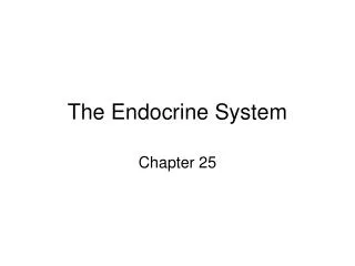
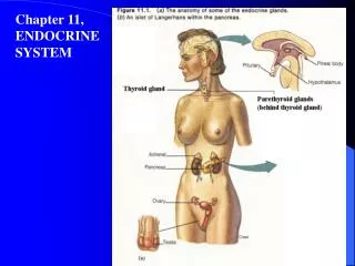
Endocrine Glands of Reproduction Body Diagram of Endocrine Glands
The reproductive process in mammals is governed by the central nervous system. Information emaniting from a variety of external cues (eg. Visual, auditory, tactile, olfactory) is fed into the centralnervous system and converges on the Hypothalamus. The information is proessed, amplified, transduced to a humoral signal and transmitted to the anterior pituitary gland where it is further amplified and transmitted via the gonadotrophic hormones to gonads. The latter respond in many ways, one of which is by the secretion of sex hormones. These, in turn, act on a host of target tissues including the brain and pituitary gland. This form a vastly complex network of information transfer, a network that permits amplification, propagation and integration of signals throughout the body. Organization of control system governing reproduction
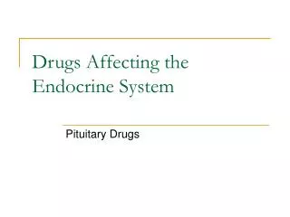
Hypothalamus The hypothalamus is located in the brain, at the base of the optic chiasm. The hypothalamus consists of nervous tissue lying inferior to the two lobes of the thalamus. The pituitary gland, or hypophysis, is found at the base of the brain below the hypothalamus and the two structures are connected via the infundibulum, or pituitary stalk, which carries both axons and blood vessels. It secretes hormones that stimulate or suppress the release of hormones in the pituitary gland, in addition to controlling water balance, sleep, temperature, appetite, and blood pressure Brain Diagram of Endocrine Glands
The hypothalamic boundary is limitted frontally by the optic chiasma, caudally by the mamillary bodies and dorsally by the thalamus. The hypothalamus surrounds the fluid-filled third ventricle. It is made up of various types of structural elements including cell bodies of hypothalamic neurons with their axons and terminals. Axons and terminals of other neurons with cell bodies lying outside the hypothalamus and axons passing through from extrahypothalamic neurons. The cell bodies of the hypothalamic nuclei send axonal projections to one of four general regions : Other areas of the brain Other hypothalamic nuclei The median eminence The posterior lobe of the pituitary gland Anatomical organization of the Hypothalamo-hypophyseal axis
The median eminence The median eminence comprises the base of the hypothalamus and is continuous with the pituitary stalk. It contains few, if any, nerve cell bodies, but consists of axons and terminals of both hypothalamic and extrahypothalamic neurons, glial cells and specialized ependymal cells called tanacytes. The latter cells line the third ventricle and are of potential importance in the transfer of information from the cerebrospinal fluid to the pituitary gland. The neural structures of the median eminence contains a capillary plexus connected with the hypothalamo-pituitary portal system.
The pituitary gland: • The pituitary gland or hypophysis is sometimes called the "master" gland of the endocrine system, because it controls the functions of the other endocrine glands. • The pituitary gland is located at the base of the brain. The gland is attached to the hypothalumus (a part of the brain that affects the pituitary gland) by nerve fibers. • The pituitary gland itself consists of three sections: • the anterior lobe (adenohypophysis) • the intermediate lobe • the posterior lobe (neurohypophysis)
The anterior pituitary is derived from oral epithelium from the roof of the mouth cavity, which migrates upwards towards the neural tube • It sits in the sella turcica which is a depression of the sphenoid bone at the base of the skull and lies behind the sphenoid sinus. • The anterior lobe of the pituitary is further subdivided into the pars distalis, pars intermedia and pars tuberalis • The anterior pituitary makes up 75% of the total weight of the pituitary. • The pars distalis forms the major part of the gland. • The pars tuberalis surrounds the infundibular stem like a cuff and extends upwards to lie beneath a portion of the median eminence • The anterior pituitary contains no nerve fibers and terminals and so is not in direct neuronal contact with the hypothalamus. • The anterior pituitary connected to the brain by a vascular connection, the hypothalamo-hypophyseal portal system. • The anterior pituitary gland consist of many different cell types classified on the basis of their size, shape and histological staining characteristics.
Somatotrophs secrete growth hormone (GH), are distributed laterally and make up 50% of hormone secreting cells. • Thyrotrophs secrete thyrotropin (TSH), are concentrated laterally and make up 10% of cells. • Gonadotrophs secrete luteinizing hormone (LH) and follicle stimulating hormone (FSH), are randomly distributed and make up 10% of cells. • Corticotrophs secrete adrenocorticotrophic hormone (ACTH), b-lipotrophin, a -melanocyte stimulating hormone and b-endorphin, are found in the median portion and make up 15-20% of cells. • Lactotrophs secrete prolactin, are randomly distributed and make up 25% of cells. The number of these cells increases in response to increased oestrogen in pregnancy and lactation. • The posterior pituitary develops as an extension of the hypothalamus itself. • The infundibulum is formed from the neuroectoderm of the floor of the third ventricle and develops to form the posterior pituitary. • The median eminance is also formed from neuroectoderm.
anterior lobe: • growth hormone • prolactin - to stimulate milk production after giving birth • ACTH (adrenocorticotropic hormone) - to stimulate the adrenal glands • TSH (thyroid-stimulating hormone) - to stimulate the thyroid gland • FSH (follicle-stimulating hormone) - to stimulate the ovaries and testes • LH (luteinizing hormone) - to stimulate the ovaries or testes intermediate lobe: • melanocyte-stimulating hormone - to control skin pigmentation posterior lobe: • ADH (antidiuretic hormone) - to increase absorption of water into the bloodby the kidneys • oxytocin - to contract the uterus during childbirth and stimulate milk production
Blood Flow The pituitary receives blood supply from paired superior and inferior hypophyseal arteries which originate from the internal carotid arteries. The superior hypophyseal arteries enter the primary capillary plexus of the median eminance close to the nerve endings of the neuroendocrine cells of the hypothalamus. From here, blood flows down the portal veins to the sinusoidal vessels of the anterior pituitary This provides a direct vascular link between the hypothalamus and anterior pituitary secretory cells. There is also thought to be a reverse flow of peptides along the infundibulum back to the brain which may provide a rapid feedback mechanism. Venous drainage is into the dural sinuses surrounding the pituitary gland. Blood supply to the posterior pituitary is from the inferior hypophyseal arteries directly from the systemic blood flow. HYPOTHALAMO-PITUITARY AXIS STRUCTURE AND DEVELOPMENT
Nerve Pathways The hypothalamus is innervated from many areas of the brain, including the limbic system, cerebral cortex, thalamus and other areas. This is due to its important role in regulating many vital functions such as maintenance of body temperature, appetite, emotions, pain, intellect etc.as well as control of endocrine functions. The hypothalamus contains two groups of nuclei with neuroendocrine functions. These are the paired supraoptic and paraventricular nuclei and the hypothalamic-hypophyseotropic nuclei. The posterior pituitary contains the termination of axons from the supraoptic and paraventricular nuclei, which abut onto capillaries. These cells produce vasopressin and oxytocin, which are stored in and secreted from the posterior pituitary gland. The newly synthesised hormones are packaged with the protein neurophysin and move down the nerve fibres by fast axonal transport. They then move by exocytosis into the efferent blood vessels of the posterior pituitary. Some of the axons of the supraoptic nuclei terminate in the upper infundibulum so some vasopressin may still be produced following loss of the posterior pituitary.
The posterior pituitary is derived from the forebrain during development and is composed predominantly of neural tissue. The posterior pituitary lies below the hypothalamus, with which it forms a structural and functional unit: the neurohypophysis. The neurohypophysis consists of three parts: the supraoptic and paraventricular nucleii of the hypothalamus (containing the cell bodies of the magnocellular, neurosecretory neurones that synthesize and secrete VP and OT); the supraoptico-hypophyseal tract (which includes the axons of these neurones); and the posterior pituitary (where the axons terminate on capillaries of the inferior hypophyseal artery). The supraoptic nucleus (SON) is situated along the proximal part of the optic tract. It consists of the cell bodies of discrete vasopressinergic and oxytotic magnocellular neurons projecting to the posterior pituitary along the supraoptico-hypophyseal tract. The paraventricular nucleus (PVN) also contains discrete vasopressinergic and oxytotic magnocellular neurons projecting to the posterior pituitary along the supraoptico-hypophyseal tract. The PVN contains additional, smaller parvicellular neurons projecting to the median eminence and additional extra-hypothalamic areas including forebrain, brain stem, and spinal cord. Some of these parvicellular neurons are vasopressinergic. The anatomy of the Neurohypophysis
A group of those projecting via the median eminence co-secrete VP and corticotrophin releasing hormone (CRH), and terminate in the hypophyseal-portal bed of the anterior pituitary. These neurons have a role in the regulation of adrenocorticotrophin (ACTH) release. The posterior pituitary receives an arterial blood supply from the inferior hypophyseal artery and the artery of the trabecula (a branch of the superior hypohyseal artery), derivatives of the internal carotid artery and its branches. The SON and PVN receive an arterial supply from the suprahypophyseal, anterior communicating, anterior cerebral, posterior communicating and posterior cerebral arteries, all derived from the circle of Willis. Venous drainage of the neurohyphysis is via the dural, cavernous and inferior petrosal sinuses. Schematic representation of the anatomy of the neurohypophysis, and it's major afferent and efferent connections
The pineal gland is a small organ shaped like a pine cone (hence its name). It is located on the midline, attached to the posterior end of the roof of the third ventricle in the brain. The pineal varies in size among species; in humans it is roughly 1 cm in length, whereas in dogs it is only about 1 mm long. To observe the pineal, reflect the cerebral hemispheres laterally and look for a small grayish bump in front of the cerebellum. Histologically, the pineal is composed of "pinealocytes" and glial cells. Anatomy of the Pineal Gland
How does the retina transmit information about light-dark exposure to the pineal gland? • Light exposure to the retina is first relayed to the suprachiasmatic nucleus of the hypothalamus, an area of the brain well known to coordinate biological clock signals. • Fibers from the hypothalamus descend to the spinal cord and ultimately project to the superior cervical ganglia, from which post-ganglionic neurons ascend back to the pineal gland. • Thus, the pineal is similar to the adrenal medulla in the sense that it transduces signals from the sympathetic nervous system into a hormonal signal.
OVARY Structure: • A flattened ovoid shape, 3 - 5 cm in diameter • The outer (periphery) ovary is the cortex and is fibrotic. It contains: • Germ cells, or ova, which are contained in follicles • Corpora lutea - produce oestrogens and progesterones • Corpora albicantes - degenerate • The stroma is the body of the ovary, containing: • Fine collagen fibres • Scattered bundles of smooth muscle cells • Some stromal cells may contain lipid droplets • The mature ovary will contain many follicle of different sizes and development.
TESTIS Macrostructure: • Paired, ovoid structures • Capsule: • External: tunica vaginalis • Internal: tunica albuginea • Gland is divided into intercommunicating, testicular lobules by fibrous septa • produce androgen-binding globulin: • this is in response to FSH • similar to plasma sex hormone-binding globulin • maintains high androgen levels in the testis and seminal fluid • produce inhibin • inhibits FSH secretion • produce seminiferous tubule fluid • Between the seminiferous tubules are the interstitial LEYDIG CELLS where androgens are produced. • these cells only have LH receptors, which stimulate steroidogenesis • Also produce small amounts of oxytocin, endorphins, angiotensins, and prostaglandins • Microstructure: • Each testicular lobule contains seminiferous lobules - site of spermatogenesis • The basement membrane of the seminiferous tubules is lined with SERTOLI CELLS. These: • produce mullerian-inhibiting substance • convert androgens to oestrogen • act as a supportive cell for the developing germ cells (sperm) • form a blood-testis barrier
DEFINITION OF HORMONE A chemical substance secreted by an endocrine gland or group of endocrine cells that acts to control or regulate specific physiological processes, including growth, metabolism, and reproduction. Most hormones are secreted by endocrine cells in one part of the body and then transported by the blood to their target site of action in another part, though some hormones act only in the region in which they are secreted. Chemical types of hormones: Peptide - Few - Several amino acids Protein - Long chains of amino acids Glycoprotein - Protein hormone with carbohydrate molecules
HORMONE ACTION PROTEIN HORMONE STEROID HORMONE