Download
1 / 36
360 likes | 485 Vues
Biliary System and Liver. 1 23 2014. Liver. Largest gland of body 2nd largest organ What is the 1 st ? Skin How much does it weigh? Approx. 3 lbs. Liver is only internal human organ capable of natural regeneration of lost tissue!

E N D
Biliary System and Liver 1 23 2014
Liver Largest gland of body 2nd largest organ What is the 1st ? Skin How much does it weigh? Approx. 3 lbs
Liver is only internal human organ capable of natural regeneration of lost tissue! as little as 25% of a liver can regenerate into a whole liver Not true regeneration! lobes removed do not regrow- function is restored, but not original form (aka: compensatory growth) (in true regeneration, both original function and form are restored)
Falciform ligament divides liver into: 2 major lobes: Right lobe Left lobe 2 minor lobes: Caudate lobe- part of right lobe -posterior Quadrate lobe - part of right lobe -inferior
Functions of liver Main function -formation of bile Maintain a proper level or glucose in blood Convert glucose to glycogen Produce urea Make certain amino acids Filter harmful substances from blood (alcohol) Store vitamins and minerals Produce 80% of cholesterol
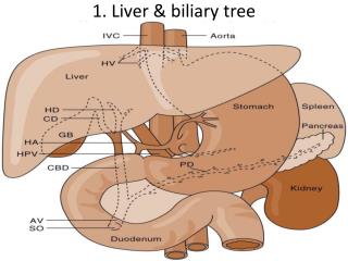
What is unique about liver? • It has a dual blood supply! • Receives both oxygenated and • deoxygenated blood (portal system) • 1. Hepatic artery- • supplies liver with oxygenated blood from • abdominal aorta to like any other part of body • 2. Portal vein- • carries deoxygenated blood from digestive • organs to be modified and filtered by liver • blood then returns to heart (by hepatic veins) • and is circulated to rest of body
First Pass Effect Problem Many drugs taken orally are substantially metabolized by portal system of liver before reaching general circulation Known as “first pass effect” Thus certain drugs can only be taken via certain other routes! suppository intravenously intramuscularly aerosol inhalation sublingually Nitroglycerin cannot be swallowed - liver would inactivate medication -must be taken under tongue or transdermally
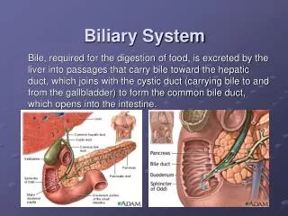
Biliary System (Excretory system of liver) Consists basically of : 1. gallbladder 2. bile ducts
Biliary Combining Forms chole – relationship with bile (aka: gall) bladder – sac or bag serving as receptacle for a secretion cyst – closed sac having distinct membrane and division with nearby tissue (May contain air, fluids, or semi-solid material) docho – duct – tube or passage way for conducting a substance angio - vessel graph- representation of a set of objects -iasis–presence of -itis – inflammation of
2 Primary Functions of Biliary System Aid in digestion- by controlling release of bile (Bile - greenish-yellow fluid produced in liver (consisting of waste products, cholesterol, and bile salts) (when excreted gives feces dark brown color) Drain waste products from liver into duodenum
Gall bladder Reservoir for bile from liver – 2oz. capacity (50 percent of bile is stored in gallbladder) Concentrates bile How much bile does it produce per day? 1-3 pints How does bile get into gallbladder? Sphincter of Oddi closes up, and bile is re-routed up into GB for temporary storage when not needed
When food containing fat enters digestive tract… the release of bile from the gallbladder is stimulated by secretion of a hormone called cholecystokinin
Transportation of bile sequence Liver secretes bile- into right and left hepatic ducts which join to become common hepatic duct which joins with cystic duct from gallbladder to become the: common bile duct which joins with pancreatic duct to form a junction known as: hepatopancreaticampulla (or ampulla of vater Spincter of Oddi (or spincter of hepatopancreaticampulla)controls emptying of bile into duodenum
Gallstones Hardened deposits of digestive fluid that can form in gallbladder Range in size from grain of sand to Can have one or hundreds! 1 in 10 people have gallstones (can’t see if not calcified!)
Two types of gallstones 80% are cholesterol stones: usually yellow-green and made primarily of hardened cholesterol 20% are pigment stones: small, dark stones made of bilirubin
Risk Factors for Gallstones Female Age 60 or older American Indian or Mexican heritage Overweight or obese Pregnant Eating a high-fat, high-cholesterol, or low fiber diet Family history of gallstones Diabetes Losing weight very quickly Taking cholesterol-lowering medications Taking medications containing estrogen (such as hormone therapy drugs)
Complications from Gallbladder Stones Choledocholithiasis - presence of bile stones in ducts Cholecystitis - bile sac inflammation Pancreatitis Increased risk of gallbladder cancer (very rare)
Treatment for Gallstones Surgical removal of gallbladder - Cholecystectomy Use medicines to dissolve stones (isn't suitable for everyone -may take a very long time) Shock-wave lithotripsy ( high-energy sound waves) to break gallstones into tiny fragments, then dissolved by medicines
If your gallbladder is removed… No longer a holding space to store bile Bile continuously runs out of liver, through the hepatic ducts, into common bile duct, and directly into small intestine When a high-fat meal is eaten - not enough bile available to digest it properly Can result in chronic diarrhea Small intestine’s ability to absorb essential fatty acids, vitamins and minerals is compromised without help of gallbladder
Pancreas Both an exocrine and endocrine gland! Endocrine- (Isle of Langerhans) produces glucagon and insulin to regulate sugar metabolism Exocrine- secretes digestive enzymes Generally cannot be seen on radiographs
Radiological exams of Gallbladder (largely replaced by Ultrsound, CT, MRI, nuclear medicine) Cholecystography Study of gallbladder Oral contrast is used Cholangiography Study of biliary ducts IV contrast is used (may be injected directly into ducts)
Cholelithiasis (gallstones) -bile calculi presence Cholecystitis (inflammation of gallbladder)-bile sac inflammation Check liver function Biliaryneoplasia(tumor or mass in biliary system) Biliarystenosis(abnormal narrowing of ducts) Demonstrate concentrating/emptying ability of gallbladder IndicationsforBiliary Tract Exam
Contraindicationsfor performing Biliary Tract Exams • Allergy to contrast • Pyloric obstruction (blockage from stomach to duodenum) • Severe jaundice • Malabsorption • Liver dysfunction • Hepatocellular disease- liver typically inflamed and shows signs of injury
Patient Prep Fat-free meal evening before Oral contrast taken 2 to 3 hours after evening meal NPO after midnight until exam Avoid laxitaves less than 24 hours to avoid prevent voiding of contrast medium with fecal material Make sure patient can, will, and did follow instructions! Early morning appointment
Position of Gallbladder • RUQ • In hypersthenic pt. • Superior and lateral • In Asthenic • Inferior and nearer to spine
ShieldingWhat 3 things must you consider? 1. Are gonads within 2” of primary x-ray field after proper collimation? 2. Are clinical objectives compromised? 3. Does pt have reasonable reproductive potential?
Gallbladder Exam(Cholecystography) Scout film will also demonstrate if contrast is visible in gallbladder Dr. may do fluoroscopic examination Post-fatty meal film may be obtained to demonstrate emptying ability of GB
PA Projection Patient prone- or upright facing wallboard Center 10x12 cassette at RUQ, level of the right elbow 70 - 80 kVp range Exposure made at end of full? expiration
PA Oblique Projection LAO position Pt rotated 15 - 40 degrees depending on body habitus CR at level of elbow, between spine and (R or L?) midaxillary line 10x12 cassette
Rt. Lateral Decubitus Demonstrates stones lighter than bile visible only by stratification CR: Directed horizontally to level of gallbladder
Very rarely performed anymore Used when patients cannot tolerate oral contrast Generally done in supine, and RPO positions Films taken at timed intervals - up to about 40 minutes after injection Intravenous Cholangiography (IVC)
Percutaneous Transhepatic Cholangiography(performed preoperatively) (Percutaneous: any medical procedure where access to inner organs or other tissue is done via needle-puncture of skin, rather than by scapel) Long needle (Chiba) is placed into bile ducts Contrast is injected under fluoro Biliary drainage or stone extraction may accompany this procedure
Cholangiography Intra-operative Performed during a cholecystectomy Examines patency of ducts during or after surgical removal of GB
T-Tube Cholangiography Post-operative (after cholecystectomy) procedure performed through T-tube left in common hepatic and common bile ducts (for drainage) To determine: patency (openness) of biliary ducts after cholecystectomy status of Spincter of oddi presence of residual or undetected stones
3 Cholangiogram types compared Percutaneous Intraoperative T-Tube
ERCP Endoscopic Retrograde Cholangiopancreatography Used to diagnose biliary and pancreatic pathologic conditions when ducts are not dilated and ampulla is not obstructed Fiberoptic endoscope passed through mouth into duodenum under fluoroscopy Common bile duct is catheterized Contrast is injected