Download
1 / 39
440 likes | 871 Vues
Hereditary Hemochromatosis. Our Patient . 55 year old white male Chief complaint: “Really tired all the time.” . History and Physical. Pt complains of extreme fatigue for the past six months. Pt has less endurance for his favorite activities.

E N D
Our Patient • 55 year old white male • Chief complaint: “Really tired all the time.”
History and Physical • Pt complains of extreme fatigue for the past six months. • Pt has less endurance for his favorite activities. • Pt feels that his exercise tolerance is decreased – he gets short of breath easily. • Pt cannot identify exacerbating or relieving factors. • Associated symptoms: • Abdominal fullness. • Severe joint pains. • Frequent urination. • Thinks he may get occasional leg edema, but isn’t sure. • Occasional heart palpitations. • No fevers.
Differential Diagnosis of Subacute Fatigue • Broad differential diagnosis! • Cardiac disease • Renal failure • Liver failure • Endocrine disease • Thyroid? • Adrenal? • HPA axis? • Autoimmune disease • Chronic infection • Bacterial? • Viral? • Fungal? • Depression • Cancer • What do you want to do for this patient?
History and Physical, Continued. • PMH: • Hypertension, past 5 years. Well controlled on thiazide. • Arthritis, past 15 years in hips, knees, hands. • Low testosterone, past 5 years. • PSH: • None.
History and Physical, Continued. • FH: • Mother is alive at 80; has mild hypertension and Alzheimer dementia. • Father died of congestive heart failure at 59. Had hypertension, diabetes mellitus, and severe arthritis. • Siblings: • Sister, 60, recently diagnosed with diabetes mellitus. • Brother, 57, healthy. • Brother, 52, advanced alcoholic cirrhosis. • Sister, 49 and healthy. • Pt has two healthy children, ages 22 and 24.
History and Physical, Continued. • Social History. Pt is married. Nonsmoker and nondrinker. Worked as a business executive until he retired this year due to symptoms. • Review of systems, significant points. • Skin: Increased pigmentation; denies sun exposure. Pt notices that his breast tissue has grown. • Cardiovascular: Worsening exertionaldyspnea over the past three years. Can climb 2 flights of stairs. Has had 2-pillow orthopnea for the past 6 months. No syncope or palpitations. • Hematologic: Pt notices that he bruises more easily. • Endocrine: Decreased sexual drive. • Musculoskeletal: Worsening arthritis.
Initial Labs • Chem 10 • Na+, K+, HCO-3, Cl-, BUN, Creatinine, Glucose, Ca++, Mg++, PO4. • CBC with differential • Serum liver enzymes (AST, ALT, AlkPhos) • Serum B-type Natriuretic Peptide • Serum TSH and total T4. • Serum Iron studies
Lab Results: • WHICH OF THESE ARE ABNORMAL? • Na+: 140 mEq/L • K+: 4.0 mEq/L • Cl-: 100 mEq/L • HCO3: 24 mEq/L • BUN: 30 mg/dL • Cr: 1.0 mg/dL • Glucose: 240 mg/dL • Ca++: 9.8 mg/dL • Mg++: 2.0 mg/dL • PO4: 3.0 mg/dL
Lab Results: • WHICH OF THESE ARE ABNORMAL? (Abnormals are in red). • Na+: 140 mEq/L (normal ≈ 137-145 mEq/L) • K+: 4.0 mEq/L (normal ≈ 3.5-5.0 mEq/L ) • Cl-: 100 mEq/L (normal ≈ 98-110 mEq/L ) • HCO3: 24 mEq/L (normal ≈ 22-26 mEq/L ) • BUN: 18 mg/dL(normal ≈ 7-21 mg/dL ) • Cr: 1.0 mg/dL(normal ≈ 0.5-1.4 mg/dL ) • Glucose: 240 mg/dL random fasting Abnormal. (Normal ≈ 65-110mg/dL) • Ca++: 9.8 mg/dL(normal ≈ 8.5-10.4 mg/dL ) • Mg++: 2.0 mg/dL(normal ≈ 1.5-2.5 mg/dL ) • PO4: 3.0 mg/dL(normal ≈ 2.5-4.5 mg/dL )
Lab Results: • Which of these is abnormal? • CBC with differential: • Hemoglobin: 15 g/dL • Hematocrit: 45% • Plt: 357,000/μL • WBC: 8,500/μL; 74% Neutrophils, 24% Lymphs, 1% Eosinophils, 1% Basophils
Lab Results: • Which of these is abnormal? • Answer: None. All are OK. • CBC with differential: • Hemoglobin: 15 g/dL(Normal male ≈ 13.2-16.2 g/dL) • Hematocrit: 45% (Normal male ≈ 40-52%) • Plt: 357,000/μL (Normal ≈ 140,000-350,000/µL) • WBC: 8,500/μL; 70% Neutrophils, 24% Lymphs, 4% Monocytes; 1% Eosinophils, 1% Basophils(Normal ≈ 4,100-10,900 WBC; Neutrophils 35-80%; Lymphocytes 20-50%; Monocytes 2-12%; Eosinophils 0-7%; Basophils 0-2%)
Lab Results • Which of these are abnormal? • Liver Enzymes • AST: 201 U/L • ALT: 186 U/L • AlkPhos: 314 U/L • B-type Natriuretic Peptide: 450 pg/mL • Endocrine • TSH: 0.1µU/mL • Total T4: 1.0 mg/dL • Free T4 0.2 mg/dL
Lab Results? • Which of these are abnormal? • Liver Enzymes • AST: 150 U/L (Normal ≈ 5-35 U/L) • ALT: 107 U/L (Normal ≈ 7-56 U/L) • AlkPhos: 214 U/L (Normal ≈ 38-126 U/L) • B-type Natriuretic Peptide: 450 pg/mL(<100pg/mL is normal; 100-300pg/mL suggests CHF; 300-600pg/mL indicates mild CHF) • Endocrine • TSH: 0.1 µU/mL (Normal ≈ 0.4-4.0µU/mL) • Total T4 1.0 mg/dL(Normal ≈ 4.5-11.5 mg/dL) • Free T4 0.2 mg/dL(Normal ≈ 0.7-2.0 mg/dL)
Lab Results • Which of these are abnormal? • Iron Studies • Serum Iron Concentration: 250µg/dL • Serum Ferritin: 3,000ng/mL • Transferrin Saturation: 80% • Total Iron Binding Capacity: 200µg/dL
Lab Results • Which of these are abnormal? • Iron Studies • Serum Iron Concentration: 250µg/dL • (Normal male ≈ 50-150 µg/dL) • Serum Ferritin: 3,000ng/mL • (Normal male ≈ 20-300ng/mL) • Transferrin Saturation: 80% • (Normal ≈ 14-50%) • Total Iron Binding Capacity: 200µg/dL • (Normal ≈ 250-450 µg/dL)
Lab Results Recap • Elevated Liver Enzymes • Hypothyroidism (low TSH and T4) • Mild Congestive Heart Failure • Elevated Blood Glucose • Serum Iron Studies suggest Iron Overload. • What will we do to confirm the suspected diagnosis? • What other tests do we want?
Next Steps? • Liver • Biopsy • Cardiac Evaluation • Cardiac Imaging Options • Electrocardiogram • Biopsy?
Liver Biopsy Results No cirrhosis noted. Hemosiderin granules inside hepatocytes. Note the bright staining with Prussian Blue. Hemosiderin granules at lower-power magnification, without Prussian blue stain.
Cardiac Studies • Echocardiogram – shows biventricular diastolic dysfunction. • Holter EKG – shows atrialtachyarrhythmias. • Myocardial biopsy – shows hemosiderosis (intracellular iron). Surviving myocytes
Hereditary Hemochromatosis • Autosomal Recessive • Associated with mutations in the HFE gene, whose protein product modulates iron transport from duodenal enterocytes into the bloodstream. • Affects 8/1000 of people of Northern European descent; as many as 10% of U.S. whites may be carriers. • 50% of affected males and 25% of affected females suffer serious complications. • The most common disease-associated allele is the C282Y mutation; an H63D mutation is also reported. Compound heterozygotes may display a disease phenotype. • Treatment is therapeutic serial phlebotomy. Even if instituted late, this can improve patient symptoms, most notably cardiac ones.
Pathophysiology • With insufficient HFE function, inappropriate amounts of iron accumulate in the body. • When found intracellularly, this iron is referred to as “hemosiderin.” At left, panels A-D, hemosiderin granules in liver tissue. Note the intracellular location of the granules. Prussian blue stain
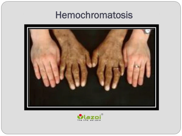
Pathophysiology, Continued • Hemosiderin deposition is thought to cause tissue damage by generating free radicals. • Hemosiderin may deposit in the anterior pituitary. • Hemosiderin may deposit in the liver. • Hemosiderin may deposit in the pancreas. • Hemosiderin may deposit in the myocardium. • Hemosiderin may deposit in joints. • May present as severe, diffuse osteoarthritis. • Hemosiderin may deposit in the skin. • Patients, who are often naturally light-skinned, may experience an unexplained darkening of their skin.
Hereditary Hemochromatosis – Clinical Features • Affected males – symptoms usually manifest after age 40. • Affected females – symptoms usually manifest several years after menopause. • This disparity is thought to be related to females’ tendency to lose iron through menses and in childbearing. • The “classic” presentation is that of “bronze diabetes”, or a constellation of symptoms including skin hyperpigmentation and diabetes mellitus; this represents a very late stage of the disease. • Today, it tends to present earlier; the most common presenting symptoms are nonspecific, and include fatigue, abdominal pain, and arthritis.
Hereditary Hemochromatosis – Clinical Features • LIVER: • Iron overload eventually leads to hepatic inflammation, fibrosis, and cirrhosis. • May initially manifest as hepatomegaly or abdominal discomfort. • Patients frequently present with elevated liver enzymes, or liver failure. • Patients are more susceptible to alcoholic cirrhosis, and should be counseled to avoid alcohol.
Hereditary Hemochromatosis – Clinical Features • Hypothalamic-Pituitary Axis. • Hemosiderin deposits in the anterior pituitary. • In men, loss of gonadotropins may result in low libido, testicular atrophy, and gynecomastia. • In all patients, TSH levels may be low, resulting in hypothyroidism. • The thyroid gland itself may be affected by hemosiderin deposition too; this may lead to transient hyperthyroidism followed by hypothyroidism.
Hereditary Hemochromatosis – Clinical Features • Pancreas • Hemosiderin deposits in the pancreas, leading to eventual endocrine pancreatic failure. • End result is diabetes mellitus.
Hereditary Hemochromatosis – Clinical Features • CARDIAC • Hemosiderin deposits in myocardial cells. • Eventually results in diastolic dysfunction; clinically presents as a restrictive cardiomyopathy. • In severe cases, clinical picture may be one of congestive heart failure. • Increased incidence of atrial arrhythmias in these patients. • Heart blocks may occur.
Hereditary Hemochromatosis – Clinical Features • Immunologic: Unusual infections • Due to their high serum iron levels, hereditary hemochromatosis patients are susceptible to severe systemic infections by certain pathogens which can include... • Listeriamonocytogenes– found in deli meats, cheese, unwashed fruits. • Vibriovulnificus – found in raw seafood and salt water. • Yersiniapseudotuberculosis – foodborne or zoonotic. • Rhizopusorayzae – virulent, angioinvasive fungus.
Hereditary Hemochromatosis – Clinical Features • Ophthalmologic: Angioid streaks. • Represents small breaks in Bruch’s membrane, in the choroid layer of the eye. • Found in HH as well as other metabolic diseases. • Patients are prone to developing neovascularization and retinal damage. • Patients should be advised to wear polycarbonate safety glasses.
Radiographic Correlates Liver is more opaque than normal. CT scan of a patient with advanced hereditary hemochromatosis. Enlarged spleen suggests that this patient has portal hypertension secondary to liver damage.
Radiographic Correlates Normal liver. Note the uniform attenuation pattern of the liver, and how it has about the same density as the spleen. Hemochromatosis. Note how the liver is hyperattenuating compared with the spleen. Also note that the spleen is enlarged.
Hemochromatosis and Arthritis Chondrocalcinosis, or the deposition of calcium pyrophosphate crystals in the articular cartilage, can occur in hereditary hemochromatosis.
Hemochromatosis and Arthritis Serial hand radiographs of an HH patient. Subchondral sclerosis (intense white band) Joint space narrowing. Fairly normal radiograph. 10 years later. Note the preserved joint spaces and the relatively uniform density of the bones.
Other Histologic Findings in Hereditary Hemochromatosis This pancreas has extensive deposition of hemosiderin granules in the acinar cells.
Hereditary Hemochromatosis - Treatment • The mainstay of treatment is serial phlebotomy. • May prevent onset of severe symptoms in asymptomatic patients. • May lead to reversal of cardiomyopathy. • Cirrhosis and pancreatic dysfunction are irreversible.
Screening for Hereditary Hemochromatosis • No consensus on general population screening. • Serum transferrin saturation is preferred by the AAFP, but false positives may occur in chronic hepatitis and alcoholic liver disease. • Ferritin is very sensitive, but extremely nonspecific. • Screen first-degree relatives of patients. • HFE gene analysis is preferred, because of the poor specificity of other noninvasive tests.
Resolution • The patient began a regimen of serial phlebotomy, and his serum iron levels improved. • Patient’s liver enzyme levels returned to near normal. Luckily, he was treated before the onset of cirrhosis. • Patient’s cardiac function improved as well. • Patient continued to suffer from arthritis and required bilateral knee replacements five years later. • Patient will require diabetes care and thyroid hormone replacement for the rest of his life. • Patient avoids alcohol, due to his increased risk for cirrhosis. • Patient avoids seafood and sea water, due to his increased risk for Vibriovulnificusinfection.
Resolution • Patient’s family was screened for hereditary hemochromatosis. • Review of patient’s father’s medical records raises suspicion that he was affected as well. • Pt’s older sister and younger brother were found to have evidence of symptomatic hemochromatosis as well. • Pt’s son had elevated serum iron levels, and was counseled about the need for therapeutic phlebotomy.