Download
1 / 17
190 likes | 378 Vues
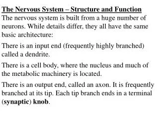
PSYC2001 Physiological Basis of Behaviour Lecturer: Associate Professor Richard Clark Lecture 2 Anatomy of the Nervous System. Anatomy of the Nervous System. Structure of the Vertebrate Nervous System. Central nervous system : the brain and the spinal cord

E N D
PSYC2001 Physiological Basis of BehaviourLecturer: Associate Professor Richard ClarkLecture 2Anatomy of the Nervous System
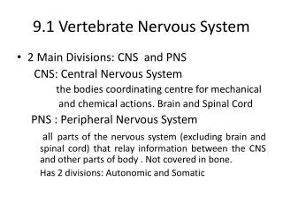
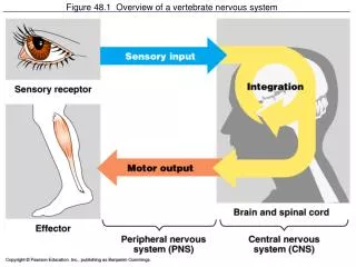
Anatomy of the Nervous System Structure of the Vertebrate Nervous System • Central nervous system: the brain and the spinal cord • Peripheral nervous system: the nerves outside the brain and spinal cord • Somatic: the nerves that convey messages from the sense organs to the CNS and from the CNS to the muscles and glands • Autonomic: a set of neurons that control the heart, the intestines, and other organs • sympathetic: arousal, “fight or flight”, emergency • parasympathetic: “relax and digest”, non-emergency
Anatomy of the Nervous System Spinal cord (CNS) and somatic PNS interface Diagram of a cross section through the spinal cord. The dorsal root on each side conveys sensory information to the spinal cord; the ventral root conveys motor commands to the muscles. Cut the spinal cord and brain loses motor control over parts of body served by that segment and below
Anatomy of the Nervous System Spinal cord (CNS) and Autonomic PNS • Sympathetic: prepares the body for arousal • breathing, heart rate, digestion • ganglia just outside spinal cord • short preganglionic and long postganglionic axons (release noradrenaline) • Parasympathetic: facilitates vegetative, non emergency responses by the body’s organs • increases digestive activity, activities opposing sympathetic system • consists of cranial nerves and nerves from sacral spinal cord • long preganglionic axons extend from the spinal cord to parasympathetic ganglia close to each internal organ • postganglionic fibers release acetylcholine
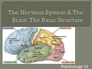
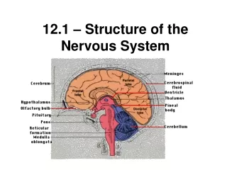
The brain - visual overview Anatomy of the Nervous System
Critical to consciousness Hind Brain Anatomy of the Nervous System Medulla oblongata Vital reflexes e.g. digestion, breathing,heart rate Damage results in vegetative (PVS) and minimal conscious (MCS) states Cerebellum Movement modulation (force, range) Learning of skills Pons Movement info. to cerebellum Critical role of mid Pons and upwards to cognitive consciousness & motor function Reticular formation (RF) Integration to modulate cortical systems arousal (ARAS) e.g. Locus coeruleus – attention (noradrenaline) Raphe nuclei - response readiness (serotonin/5-HT) Substantia nigra - movement initiation (dopamine) Also spinal motor control
Anatomy of the Nervous System Mid Brain Tegmentum Continuation of RF modulator systems Ventral tegmentum – motivation systems Raphe nuclei - response readiness (serotonin/5HT) Substantia nigra - movement initiation (dopamine) Localised damage to RF - coma Afferent and efferent fibres Tectum Superior Colliculus Low level visual processing and visuomotor integration (e.g. saccades) Inferior Colliculus Low level auditory and somatic processing; tonotopic maps; sound localisation Involvement in auditory and visual reflex control
Anatomy of the Nervous System The Diencephalon Thalamus Sensory relay station Information to sensory cortices Gating (selective attention) via nucleus reticularis thalamus (NRT) Maintains/breaks alpha (gated activity) Bilateral information flows with associative cortex e.g. Dorsomedial nuclei, prefrontal cortex and memory Hypothalamus Regulates many functions and systems (drives) Endocrine (hormonal), autonomic and visceral systems.Homeostasis, sexual behavior, fighting, feeding. Pituitary Gland Part of endocrine system Linked to hypothalamus Releases hormones into bloodstream, which carries them to other organs
Anatomy of the Nervous System The Limbic system • Critical role in learning • linked to evaluation and memory storage of significance of events and information • Integration of new with old memories via cortex • Links to Orbitofrontal cortex • Regulation of social and affective aspects of behaviour • Emotional response • fight and flight and associated aggressive behaviours • amygdala coordinates autonomic and endocrine response of emotional states
The basal ganglia and basal forebrain Anatomy of the Nervous System • Basal Ganglia • Higher level motor regulation such as movement initiation and sensorimotor integration • Aspects of motor memory and emotional expression • Basal Forebrain • N. basalis located antero-ventral to basal ganglia • Send axons to cerebral cortex • Modulatory role in memory-related arousal and attention – Acetylcholine (ACh) Nucleus basalis
Anatomy of the Nervous System Ventricular system • Key structures • Central canal: fluid-filled channel in spinal cord • Ventricles: four fluid-filled cavities within brain • Meninges: the membranes surrounding brain and spinal cord (CSF enters via 4thVent.) NB inflammation: meningitis • Cerebrospinal fluid (CSF) • Clear fluid like plasma, formed in cells of choroid plexus. Flow thru key structures • Transfers hormones from endocrine system to brain, spinal chord • Cushions the brain • Blockages: hydrocephalus; shunts
Anatomy of the Nervous System Cerebral Cortex • 10,000 million neurones • 200 million callosal projections • 5-15% sensory and motor cortex • 85% or more association cortex • Complex and multi-system: local and widely distributed neural networks • Serial and parallel architecture • Sensori motor elaboration (hierarchical) • Working memory systems (sustained neural loop activity) Sylvian fissure
Subsystems in cerebral cortex (e.g. Language) Anatomy of the Nervous System
Anatomy of the Nervous System How the Parts Work Together • Does the brain operate as a whole or a collection of parts? • Each brain area has a function but it can’t do much by itself (particularly in terms of higher function) • The Binding Problem • The question of how the visual, auditory, and other areas of your brain influence one another to produce a combined perception of the single object • Perceptual recognition produces gamma waves 30-80/second in various brain areas – creates “unified experience” • Einstein’s brain had larger than normal inferior parietal cortex, an area known to bind different aspects of perception, e.g., damage prevents binding color to shape • Critical role of working memory systems
Anatomy of the Nervous System Research Methods • Structural imaging • CT scanning: X-rays used (sometimes with dye contrasts) to take images of the brain • Magnetic Resonance Imaging (MRI): Viewing of relaxation time of hydrogen nuclei placed in strong magnetic field allows viewing of neural tissue • Trans-cranial magnetic stimulation (TCMS): intense magnetic field temporarily inactivates area • Lesion techniques (induced brain damage) • Electrode, chemicals (e.g. 6-OHDA) or excision to damage specific area • Correlates usually with behavioral impairment, e.g.,damage to Broca’s area associated with inability to speak, but may not relate directly to behavior e.g. directly affects memory, mentation
Anatomy of the Nervous System Research Methods cont. • Brain stimulation (e.g. Penfield) • can evoke sensations, e.g., flashes of light; whole experiences • less intense magnetic fields stimulate an area • injected chemicals stimulate specific receptors • but, sensations are in isolation and behaviors depend on coordination across brain areas
Anatomy of the Nervous System Research Methods Recording brain activity • Haemodynamic methods • PET (positron emission tomography) detects radioactive chemicals absorbed by most active cells. Similar to an earlier method called "regional cerebral blood flow" (rCBF). • fMRI (functional magnetic resonance imaging) detects release of oxygen in active cells - replaces PET and rCBF for measurement of general brain activity. • Electromagnetic measures (mass neural activity) • Brain electrical fields (EEG/ERPs) • Magnetoencephalography (MEG) • But, brain is always active and interpreting changing activity is a challenge; also, different people use different areas for same task