Download

1 / 33
330 likes | 351 Vues
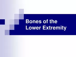
The Lower Extremity. BIOL 1010. FEMUR. TIBIA. FIBULA. ERROR: #7 is CUBOID #4 is LATERAL CUNEIFORM. Very similar to the upper extremity Some definitions: a. Thigh – part from the hips to knees b. Leg – part from the knee to the ankle

E N D
The Lower Extremity BIOL 1010
ERROR: #7 is CUBOID #4 is LATERAL CUNEIFORM
Very similar to the upper extremity • Some definitions: a. Thigh – part from the hips to knees b. Leg – part from the knee to the ankle • Note that the hip joint is a ball and socket joint. It allows movement in all direction. The knee only allows flexion and extension – not circumduction. It is never normal for hyperextension of knee (genu recurvatum)
CAT In humans, the abdominal aorta terminates into the common iliac arteries. The common iliacs divide into the external and internal iliacs. In humans, the aorta terminates as the middle sacral artery.
The nerves of the lower extremity are simpler than the upper limb. There are three nerves entering the thigh. Their lateral branches serve the gluteal muscles. femoral nerve L2,3,4 anterior compartment obturator nerve L2,3,4 medial compartment sciatic nerve L4,5,S1,2,3 (sacral plexus) posterior compartment
The only one of the three that travel below the knee is the sciatic nerve. • The sciatic nerve branches into two nerve: 1. tibial nerve 2. common peroneal nerve a. Superficial peroneal nerve b. Deep peroneal nerve
Anterior compartment- Extensors of the knee Femoral Nerve lateral medial femur Medial compartment- Adductors of the knee Obturator Nerve Posterior compartment- Flexors of the knee Sciatic Nerve
The anterior compartment of the thigh is homologous to the posterior compartment of the arm. • The posterior compartment of the thigh is homologous to the anterior compartment of the arm.
The leg has three compartments • Anterior compartment - dorsiflexors (flexors) of the foot - deep peroneal nerve • Posterior compartment - plantarflexors (extensors) of the foot - tibial nerve • Lateral compartment - extensors of the foot - superficial peroneal nerve
The FASCIA LATA (not to be confused with the muscle of similar name) is connective tissue on the lateral side of the femur. This forms the ILIOTIBIAL BAND. This band connects the ilium and tibia and is the site of inflammation in runners – iliotibial band syndrome.
Muscles of the Medial Compartment of the ThighObturator Nerve
Muscles of the Posterior Compartment of the ThighTibial portion of Sciatic Nerve
The rectus femoris cross two joints, therefore it has actions on the two joints involved. • The patella is articulates with the FEMUR, not the tibia. • The patella is attached to the femur and tibia by the PATELLAR LIGAMENT. When this ligament is pulled, the patella is lifted superiorly and brings the leg into an extended position. • In the posterior leg, the GASTROCNEMIUS muscle is responsible for plantarflexion. It originates at the femur and inserts on the posterior surface of the calcaneus. It crosses two joints so it can flex the knee and plantarflex (extend) the foot. It is innervated by the tibial nerve.
Motor Branches of the Femoral Nerve L2 L3 L4 Rectus femoris iliopsoas pectineus sartorius Vastus lateralis Vastus medialis Vastus intermedius
Motor Branches of the Obturator Nerve Adductor brevis Adductor longus gracilis Adductus magnus
Motor Branches of the Sciatic Nerve L4 L5 S1 S2 S3 Semitendinosis Biceps femoris semimembranosus Hamstrings Common peroneal nerve Tibial nerve Deep peroneal nerve Superficial peroneal nerve Anterior compartment of leg Gastrocnemius and posterior compartment of the leg and foot Lateral compartment of leg
The Foot Medial, intermediate, and lateral cuneiforms navicular talus calcaneus phalanges metatarsals cuboid
Anterior Leg Structures - Superficial Identify the Following: Tibialis Anterior Peroneus Longs Peroneus Brevis Extensor Digitorum Longus Patellar Tendon Patella Tibia Peroneus Tertius Extensor Hallucis Longus Popliteus Tendon Sartorius Tendon
Posterior Leg Structures - Superficial Identify the Following: Femur Tibia Fibula Soleus Achilles Tendon Plantaris Popliteus Calcaneus
Posterior Leg Structures - Deep Identify the Following: Achilles Tendon Flexor Hallucis Longus Flexor Digitorum Longus Calcaneus Talus Tibia Fibula Femur
Intrinsic Muscles of the Foot • These muscles all originate and insert on foot bones. • They help to flex, extend, abduct, or adduct the toes. • All the intrinsic muscles of the foot are found on the plantar surface (except the one on the dorsal aspect). • The plantar muscles are arranged in 4 layers, from superficial to deep.