Download
1 / 63
750 likes | 1.19k Vues
Principles of Radiation Protection – Managing Radiation Protection. The Management Principles. JUSTIFICATION OPTIMISATION LIMITATION. Justification.

E N D
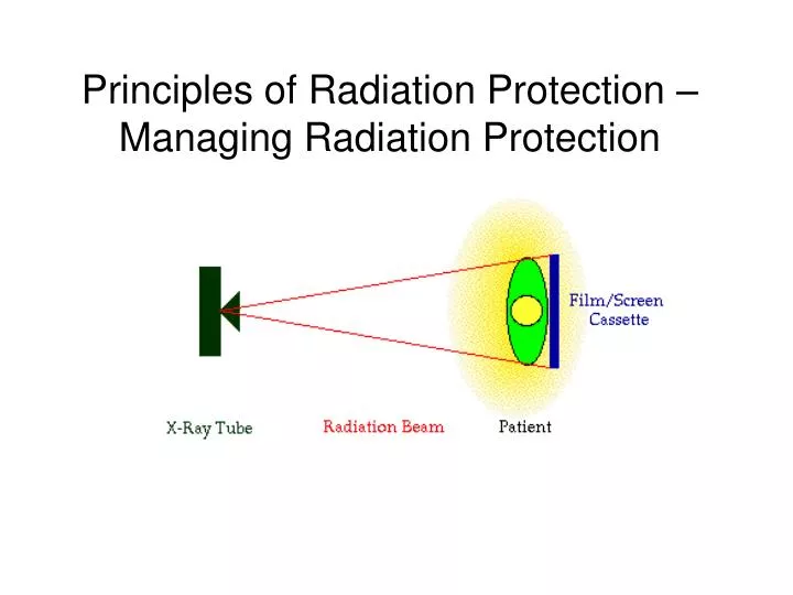
Principles of Radiation Protection – Managing Radiation Protection
The Management Principles JUSTIFICATION OPTIMISATION LIMITATION
Justification • No practice involving exposures to radiation should be adopted unless it produces sufficient benefit to the exposed individuals or to society to offset the radiation detriment it causes. • Justification of exposures is primarily the responsibility of the medical professional i.e. the Radiologist. • The expected clinical benefit associated with each type of procedure should have been demonstrated to be sufficient to offset the radiation detriment.
Justification Benefit of the radiation exposure must outweigh the risk of exposure vs
Optimisation • All exposures and radiation doses must be kept • As • Low • As • Reasonably • Practicable • With economic and social factors taken into account ALARP
OPTIMISATION For every exposure, operators must ensure that doses arising from the exposure are kept as low as reasonably practicable and consistent with the intended diagnostic purpose. THIS IS OPTIMISATION
OPTIMISATION You are defined as an IRMER Operator Your ‘optimisation’ is ensuring you leave the X-ray unit/LINAC in a safe condition fit for clinical use Handover procedure
Dose Investigation Level Once you start work with ionising radiation, you are subject to legal dose limits – 6 mSv per year for non-classified workers However, we have to define a Dose Investigation Level 1.2 mSv per year Or 0.1 mSv per month This is a level of dose that should trigger an investigation in conjunction with your RPA, and ensures that you do not receive anywhere close to the legal limit.
Limitation • In the UK, legislation stipulates annual limits for the amount of radiation that may be received by staff and members of the public • Limits are set such that deterministic effects never happen • Limits are set such that chances of stochastic effects are minimised
Legal Dose Limits - Patients • For examinations directly associated with illness – there are no dose limits
Legal Dose Limits – Radiation Workers • Radiation workers are those exposed to radiation as part of their occupation • No benefit – only risk • Two subgroups depending on level of exposure: • Classified radiation worker • Non-classified radiation worker
Legal Dose Limits – Classified Workers • Receive high levels of radiation exposure • Very unlikely for dental • Require annual health check • Compulsory dose monitoring • For classified worker • Whole body 20 mSv per year effective dose (18 years old and above) • Lens of eye 150 mSv per year equivalent dose • Skin 500 mSv per year equivalent dose • Extremities (hands and feet etc) 500 mSv per year equivalent dose
Legal Dose limits for non-classified workers (all radiation workers in this Trust) • Very unlikely you will need to be classified • You only need to be classified if you are considered to approach 3/10ths of any dose limit • Relevant dose limit for you is 6 mSv whole body effective dose • Very unlikely to exceed this per year • We use a dose constraint of 0.1 mSv per month for RT and X-ray engineers • Risk assessment usually show it is very unlikely this will be exceeded • We monitor routinely with dose badges
Legal Dose Limitation - Public • The annual dose limit for a member of the public (e.g. office worker in room next door to x-ray) • 1 mSv/yr • But we use a dose constraint of 0.3mSv/yr
RADIATION TISSUE Radiation Dose • Absorbed Dose (Jkg-1) • Amount of energy deposited per kilogram • Dose to an organ or tissue • Unit is the Gray (Gy) • DOSE TO A CERTAIN PLACE IN THE BODY • Effective Dose (Jkg-1) • This is the average dose to whole body • Unit is the Sievert (Sv) • This gives us the risk of contracting cancer of the x ray exposure • THIS IS THE OVERALL DOSE TO THE WHOLE BODY
Risks Associated with X rays • Adult Exposure (per 1 mSv) • Fatal cancer (all types) 1 in 20,000 • Fatal leukaemia 1 in 200,000 • Non fatal cancer 1 in 100,000 • Heritable effects 1 in 80,000 • Childhood exposure • Fatal cancer 1 in 10,000 • Foetal exposure • Fatal cancer to 15 years 1 in 10,000 • All cancers to 15 years 1 in 17,000 • Heritable effects 1 in 42,000
Small Risks, So why worry?... • Average effective dose for radiography ~0.5 mSv • Risk of fatal cancer only 1 in 40,000 • But, large number of patients • 40 000 000+ procedures. • Therefore, 700 patients ‘killed’ each year due to x-rays. • So: • All exposures must be JUSTIFIED. • Doses to patients, and staff, must be As Low As Reasonably Achievable (ALARA principle).
Doses in Perspective • Effective dose from natural background radiation in the UK is approximately 2.7 mSv • This is 2000 times greater than a dental exposure • This natural radiation comes from • cosmic rays, • rocks and soil, • food, • radon. • Artificial radiation comes from: • Fallout from nuclear explosions • Radioactive waste discharged from nuclear power plants • Medical and dental exposures • Occupational exposures
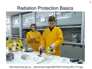
Practical methods to restrict YOUR radiation exposure • Time • Distance • Shielding
Double distance = 1/4 dose • Triple distance = 1/9th dose. In air, x-rays obey the Inverse Square Law. I∞1/d2
Distance • Operator B receives only a quarter of the radiation received by Operator A if he is standing twice the distance from the source • Operator B receives only one ninth of the radiation received by Operator A is he is standing 3 times the distance from the source
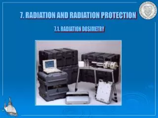
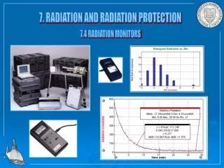
Dose monitoring • Film badges • Thermoluminescent dosemeters (TLD) • Badge • Extremities • Ionisation chambers
Film badges • Plastic frame • Worn outside of clothes for 1 to 3 months • Advantages: • Provide a permanent record of dose • Measure type and energy of radiation • Simple and robust • Disadvantages • No immediate indication of exposure • Processing can lead to errors • Prone to filter loss
TLD • Similar use as for film badges • They absorb radiation and release this as light when heated • Advantages: • Re-usable • Easy to read out • Disadvantages: • Read out is destructive • Limited info on type of radiation
Ionization chambers • Used by scientific personnel to measure beam dose • Radiation ionises air inside chamber which produces a current of electricity • Advantages • Very accurate • Immediate read out • Disadvantages • No permanent record • No indication of type of radiation • Fragile and easliy damaged
Patient Doses in Radiography - Patient Dose Limitation & Practical Principles of Radiation Protection
What are patient doses? • Absorbed dose in mGy • Dose to a certain place in the body (eg skin dose) • Effective dose in mSv • Takes into account the tissues that have been exposed
How do we measure these in practice? • Dose Area Product meters (DAP) • Skin dose: • kV, mAs, focus to skin distance • Screening time • Dose Length Product (CT Scanning)
Dose Area Product Stochastic risks approx. proportional to DAP Skin dose is DAP / area irradiated 1 Gy.cm2 3 mGy skin dose 1 Gy.cm2 0.2 mSv effective dose .
Largest Exposure from man-made radiation is Medical 46 million medical & dental x-raysin UK annually Major Contributors to UK collective dose from medical x-rays 2008 data – HPA-CRCE-012 published Dec 2010
RADIATION EXPOSURE OF THE UK POPULATION FROM MEDICAL AND DENTAL X-RAY EXAMINATIONS From NRPB/HPA data 2008 data – HPA-CRCE-012 published Dec 2010
RADIATION EXPOSURE OF THE UK POPULATION FROM MEDICAL AND DENTAL X-RAY EXAMINATIONS From NRPB/HPA data
Hundreds of cancer cases blamed on dentist x-rays Independent.co.uk By Jeremy Laurence, Health Editor Friday, 30 January 2004 Radiation from X-rays in dentist surgeries and hospitals causes 700 people in Britain to develop cancer each year, researchers say today.
700 CANCER CASES CAUSED BY X-RAYS X-RAYS used in everyday detection of diseases and broken bones are responsible for about 700 cases of cancer a year, according to the most detailed study to date. The research showed that 0.6 per cent of the 124,000 patients found to have cancer each year can attribute the disease to X-ray exposure. Diagnostic X-rays, which are used in conventional radiography and imaging techniques such as CT scans, are the largest man-made source of radiation exposure to the general population.Although such X-rays provide great benefits, it is generally accepted that their use is associated with very small increases in cancer risk. 30 January 2004
Researchers from Oxford University and Cancer Research UK estimated the size of the risk based on the number of X-rays carried out in Britain and in 14 other countries. According to their findings, published in the medical journal The Lancet, the results showed that X-rays accounted for 6 out of every 1,000 cases of cancer up to the age of 75, equivalent to 700 out of the 124,000 cases of cancer diagnosed each year.
Un-necessary exposures Those exposures that are: unlikely to be helpful to patient management, or are not As Low As is Reasonably Practicable in order to meet a clinical objective.
Factors affecting Patient Dose Field Size (Collimation) Tube voltage (kV) Beam filtration Tube to patient distance Film/Sensor speed – (Direct Digital or CR)
Collimation, Collimation, Collimation Cover only the area needed • Small fields give lower dose (and less scatter, therefore better image) • Avoid more radiosensitive areas - e.g. gonads, female breast • Position carefully.
Optimal collimation will result in : Lower patient dose Lower occupational dose Improved image quality Collimation, Collimation, Collimation