Download
1 / 44
440 likes | 566 Vues
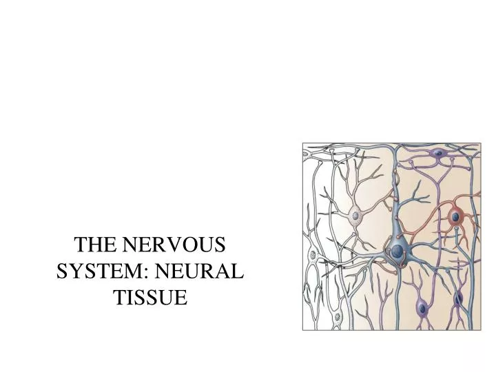
THE NERVOUS SYSTEM: NEURAL TISSUE. Nervous system functions . 1. sensory function sensory receptors detect internal and external stimuli information is sent to CNS via sensory (afferent) neurons within sensory nerves 2. integrative function

E N D
Nervous system functions • 1. sensory function • sensory receptors detect internal and external stimuli • information is sent to CNS via sensory (afferent) neurons within sensory nerves • 2. integrative function • integrates = processing of information within the CNS • stores info and also makes decisions once info is processed • one important integrative function = perception • processed by interneurons within the CNS • 90% of the neurons within the CNS are interneurons • 3. motor function • decision usually manifests itself as a motor command – contraction of a muscle, secretion by a gland • motor commands travel along motor (efferent) neurons within motor nerves • commands are sent to effectors = muscles and glands
Nervous system includes all neural tissue in body • about 3% of the total body weight • Central Nervous System • Brain and spinal cord (brain = 100 billion neurons, SC = 100 million neurons) • Peripheral Nervous System • All neural tissue outside CNS • includes the spinal and cranial nerves
A schematic of the vertebrate nervous system Figure 21-6
Cells in Nervous Tissue • Neurons • Neuroglia
Neuroglia (Glia) • about half the volume of cells in the CNS • smaller than neurons • 5 to 50 times more numerous • do NOT generate electrical impulses • divide by mitosis • Two types in the PNS • Schwann cells • Satellite cells • Four types in the CNS • Astrocytes • Oligodendrocytes • Microglia • Ependymal cells
Astrocytes • Largest of glial cells • Most numerous • Star shaped with many processes projecting from the cell body -processes make contact with the capillaries supplying CNS tissue, CNS neurons and the pia mater membrane covering the brain and spinal cord • Helpform and maintain blood-brain barrier • processes wrap around the blood capillaries and isolate the neurons from their blood supply • Provide structural support for neurons • Maintain the appropriate chemical environment for generation of nerve impulses/action potentials • regulate ion concentrations - generation of action potentials by neurons • Take up excess neurotransmitters from the synapse • Regulate nutrient concentrations for neuron survival • Assist in neuronal migration during brain development • Perform repairs to stabilize tissue – scar formation???
Oligodendrocytes • Most common glial cell type • Each forms myelin sheath around the axons of neurons in CNS • Analogous to Schwann cells of PNS • Form a supportive network around CNS neurons • fewer processes than astrocytes • round or oval cell body
Microglia • few processes • derived from mesodermal cells that also give rise to monocytes and macrophages • Small cells found near blood vessels • Phagocytic role - clear away dead cells • protect CNS from disease through phagocytosis of microbes • migrate to areas of injury where they clear away debris of injured cells - may also kill healthy cells
Ependymal Cells • epithelial cells arranged in a single layer • range in shape from cuboidal to columnar • Form epithelial membrane lining cerebral cavities (ventricles) & central canal - that contain CSF • Produce & circulate the cerebrospinal fluid (CSF) found in these chambers • CSF = colorless liquid that protects the brain and SC against chemical & physical injuries, carries oxygen, glucose and other necessary chemicals from the blood to neurons and neuroglia
PNS: Satellite Cells • Flat cells surrounding PNS axons • Support neurons in the PNS & help regulate the chemical environment surrounding the neurons • Found in the ganglia of the PNS
PNS: Schwann Cells • each cell surrounds multiple unmyelinated PNS axons with a single layer of its plasma membrane • produces part of the myelin sheath surrounding an axon in the PNS • also contributes to regeneration of PNS axons
Neurons • what is the main defining characteristic of neurons? • have the property of electrical excitability - ability to produce action potentials or impulses in response to stimuli
Representative Neuron http://www.horton.ednet.ns.ca/staff/selig/Activities/nervous/na1.htm 1. cell body or soma (or perikaryon) -single nucleus with prominent nucleolus (high synthetic activity) -Nissl bodies -rough ER & free ribosomes for protein synthesis -neurofilaments give cell shape and support –also move material inside cell -lipofuscin pigment clumps (harmless aging) - yellowish brown -the processes that emerge from the body of the neuron = nerve fibers -two kinds: dendrites & axons
Neurons 2. Cell processes = dendrites (little trees) - the receiving or input portion of the neuron -short, tapering and highly branched -surfaces specialized for contact with other neurons – either with their axons of cell bodies
3. Cell processes = axons • conduct impulses away from cell body toward synaptic end bulbs • sites for neurotransmitter storage • joins the soma at a cone-shaped elevation = axon hillock • first part of the axon = initial segment • most impulses arise at the junction of the axon hillock and initial segment = trigger zone • cytoplasm = axoplasm • plasma membrane = axolemma • side branches = collaterals arise from the axon • axon and collaterals end in fine processes called axon terminals • swollen tips called synaptic end bulbs contain vesicles filled with neurotransmitters
Axonal Transport • Cell body is the location for most protein synthesis • neurotransmitters & repair proteins • however the axon or axon terminals require neurotransmitters for signaling • Axonal transport system moves substancesto the synaptic terminals • slow axonal flow • movement of axoplasm in one direction only -- away from cell body to the terminals • only 1-5 mm per day • replenishes axoplasm in regenerating or maturing neurons • fast axonal flow • moves organelles & neurotransmitters via microtubules • at 200-400 mm per day • transports material in either direction • for transport to the terminals or for recycling things back to the cell body
Structural Classification of Neurons • can classify neurons two main ways: • 1. Structural • 2. Functional • 1. based on number of processes found on cell body • multipolar = several dendrites & one axon • most common cell type in the brain and SC • bipolar neurons = one main dendrite & one axon • found in retina, inner ear & olfactory • unipolar neurons = one process only, sensory only (touch, stretch)
Structural Classification of Neurons • Purkinje = cerebellum • Renshaw = spinal cord • 2. or can classify by the name of the histologist that first described them • 3. or can classify them by their appearance • e.g. pyramidal neurons
Functional Classification of Neurons • Sensory (afferent) neurons • transport sensory information from skin, muscles, joints, sense organs & viscera to CNS • Motor (efferent) neurons • send motor nerve impulses to muscles & glands • Interneurons (association/integrative) neurons • connect sensory to motor neurons • 90% of neurons in the body
Terms to know • membrane potential = electrical voltage difference measured across the membrane of a cell • resting membrane potential = membrane potential of a neuron measured when it is unstimulated • results from the build-up of negative ions in the cytosol along the inside of the neuron’s PM • the outside of the PM becomes more positive • this difference in charge can be measured as potential energy – measured in millivolts • polarization – change in membrane potential away from resting MP • depolarization • repolarization • hyperpolarization
ion channels in the PM of neurons and muscles contributes to their excitability • when open - ions move down their concentration gradients • two types: gated and non-gated Ion Channels 1. Leakage (non-gated) or Resting channels: are always open, contribute to the resting potential -nerve cells have more K+ than Na+ leakage channels -as a result, membrane permeability to K+ is higher -so K+ leakage is the main factor in setting the resting membrane potential • 2. Gated channels:channels can possess gates to open and close them • -open and close in response to a stimulus • A. voltage-gated: open in response to change in voltage - participate in the AP • B. ligand-gated: open & close in response to particular chemical stimuli (hormone, neurotransmitter, ion) • C. mechanically-gated: open with mechanical stimulation
Action Potential • Resting membrane potential is -70mV • AP is triggered when the membrane potential reaches and exceeds a threshold (usually -55 MV) • action potential = nerve impulse • takes place in two stages: depolarizing phase (more positive – e.g. -70 mV to -60mV) and repolarizing phase (more negative - back toward resting potential – e.g. + 30 mV to +20 mV) • followed by a hyperpolarizing phase or refractory period in which no new AP can be generated
9. The action potential http://www.blackwellpublishing.com/matthews/channel.html 8. 10. • 1. neuron is at resting membrane potential (resting MP) • 2. neuron receives a signal • Neurotransmitter (NT) • 3. NT binds ligand-gated sodium channel (LGNa) • 4. LGNa channel opens • 5. Na flows into neuron =depolarization • Inside of neuron (i.e. MP) becomes more positive • 6. if neuron depolarizes enough to Threshold = Action Potential (AP) • 7. 1st stage of AP – opening of voltage-gated Na channelsc(VGNa) • 8. even more Na flows in through VGNa channels = BIG depolarization • Membrane potential goes from negative to positive • 9. closing of VGNa channels & opening of voltage-gated K channels • 10. BIG outflow of potassium through VGK channels = repolarization • Inside of neuron (MP) becomes more negative • 11. neuron repolarizes so much – it goes past resting and hyperpolarizes • 12. closing of VGK channels • 13. all voltage-gated channels closed, Na/K pump “resets” ion distribution to resting situation 6 & 7. 4. & 5. 1. 11. 13. 12. 2 & 3.
depolarization (increase in MP) results from opening of Na+ channels. This opens an increasing number of voltage-gated Na channels which depolarizes the membrane more. Once threshold is reached, a large # of voltage-gated Na+ channels open and a rapid increase in MP results outflow of K+ restores the resting MP. Na+ channels begin to open again and K+ channels close. This results in hyperpolarization (below resting) results in a refractory period. at a certain stage of depolarization, theMP also opens voltage-gated K+ channels which permit the outflow of K+ . The Na+ channels close and the MP becomes more negative returning toward resting MP
Continuous versus Saltatory Conduction • Continuous conduction (unmyelinated fibers) • An action potential spreads (propagates) over the surface of the axolemma http://highered.mcgraw-hill.com/sites/0072437316/student_view0/chapter45/animations.html#
Saltatory Conduction • Saltatory conduction -depolarization only at nodes of Ranvier - areas along the axon that are unmyelinated and where there is a high density of voltage-gated ion channels -current carried by ions flows through extracellular fluid from node to node http://www.blackwellpublishing.com/matthews/actionp.html
Rate of Impulse Conduction • not related to stimulus strength • Properties of axon • Presence or absence of myelin sheath • myelin = 5 to 7X faster • Diameter of axon • larger = faster conduction
Action Potentials in Nerve and Muscle • another excitable cell type = muscle • muscle cells can generate their own action potential in response to the neuron’s • are some notable differences vs. Neurons • 1. entire muscle cell membrane versus only the axon of the neuron is involved • 2. resting membrane potential differ • nerve is -70mV • skeletal & cardiac muscle is closer to -90mV • 3. AP duration is longer in muscles • nerve impulse is 1/2 to 2 msec • muscle action potential lasts 1-5 msec for skeletal & 10-300msec for cardiac & smooth • BUT fastest nerve conduction velocity is 18 times faster than velocity over skeletal muscle fiber
Synapse • Synapse • Site of intercellular communication between 2 neurons or between a neuron and an effector (e.g. muscle) • Permits communication between neurons and other cells • Initiating neuron = presynaptic neuron • Receiving neuron = postsynaptic neuron • Most are axodendritic axon -> dendrite • Some are axoaxonic – axon > axon • axon terminal swell to form synaptic end bulbs • site of neurotransmitter release • multiple types of NTs can be found in one neuron type http://www.lifesci.ucsb.edu/~mcdougal/neurobehavior/modules_homework/lect3.dcr
Synapses • NTs will cause either and excitatory or inhibitory response in the post-synaptic neuron • If the NT depolarizes the postsynaptic neuron = excitatory • change in membrane potential is called an excitatory postsynaptic potential (EPSP) • produced through the opening of sodium channels or other cation channels (inward) • Some NTs will cause hyperpolarization = inhibitory • called an inhibitory postsynaptic potential (IPSP) • opening of chloride channels (inward) or potassium channels (outward) • Neural activity depends on summation of all synaptic activity • Excitatory and inhibitory
Synapses • Chemical • Membranes of pre and postsynaptic neurons do not touch • Synaptic cleft exists between the 2 neurons – 20 to 50 nm • the electrical impulse cannot travel across the cleft – indirect method is required – chemical messengers (neurotransmitters) • Most common type of synapse • The neurotransmitter induces a postsynaptic potential in the PS neuron • either an EPSP or an IPSP • Communication in one direction only http://www.blackwellpublishing.com/matthews/nmj.html
Chemical synapse • is the conversion of an electrical signal (presynaptic) into a chemical signal back into an electrical signal (postsynaptic) • 1. nerve impulse arrives at presynaptic end bulbs • 2. fusion of synaptic vesicles to PM - role for calcium in this fusion • 3. release of NTs • 4. opening of channels in PM of postsynaptic neuron (e.g. sodium) • 5. postsynaptic potential develops – possible depolarization & triggering of AP in postsynaptic neuron
Release of NTs from Synaptic end bulbs • upon arrival of the AP into the synaptic end bulbs the opening of voltage-gated Ca2+ channels • the influx of calcium promotes the “docking” of the synaptic vesicle with the PM and the exocytosis of their contents • the synaptic vesicle components are then recycled for future use Synaptic vesicles can be filled, exocytosed, and recycled within a minute
Synapses • Electrical • Direct physical contact between cells required • Conducted through gap junctions • Two advantages over chemical synapses • 1. faster communication – almost instantaneous • 2. synchronization between neurons or muscle fibers • e.g. retina, heart-beat
Neuromuscular junction Neurotransmitters • More than 100 identified • produced by neurons and stored within the neuron • secrete these NTs in response to generation of an electrical signal (action potential) by the neuron • bind onto post-synaptic neurons (synapse) or target muscle cells (neuromuscular junction) • some bind receptors on the target and cause channels to open in the target (e.g. ligand-gated sodium channels) • others bind receptors on the post-synaptic neuron and result in the opening of LG channels through other mechanisms • result in either excitation or inhibition of the post-synaptic neuron
Neurotransmitters • an important thing to consider is the removal of the neurotransmitter from the synaptic cleft • Removal of NTs • 1. Diffusion • move down concentration gradient out of the synaptic cleft • 2. Enzymatic degradation of the NT • e.g. acetylcholinesterase • 3. Uptake of the NT by neurons or glial cells (like astrocytes) • done by neurotransmitter transporters • e.g. NE, dopamine, serotonin
Neurotransmitters 1. small molecules: Acetylcholine (ACh) -All neuromuscular junctions use ACh – can only be excitatory -ACh also released at chemical synapses between two neurons -can be excitatory or inhibitory – depends on location and the neurons involved -inactivated by an enzyme acetylcholinesterase -blockage of the ACh receptors by antibodies = myasthenia gravis - autoimmune disease that destroys these receptors and progressively destroys the NMJ -anticholinesterase drugs (inhibitors of acetylcholinesterase) prevent the breakdown of ACh and raise the level that can activate the still present receptors
Neurotransmitters 2. Amino acids: glutamate & aspartate & GABA • most have powerful excitatory effects • stimulate most excitatory neurons in the CNS (about ½ the neurons in the brain) • GABA (gamma amino-butyric acid) is an inhibitory neurotransmitter for 1/3 of all brain synapses • i.e. inhibits the generation of an action potential by the target neuron - Valium is a GABA agonist - enhances its inhibitory effect
Neurotransmitters 3. Biogenic amines: modified amino acids • catecholamines:norepinephrine (NE), epinephrine, dopamine, thyroxin • all derived from the amino acid tyrosine • NE - role in arousal, awakening, deep sleep, regulating mood • epinephrine (adrenaline) - flight or fight response • dopamine - emotional responses and pleasure, decreases skeletal muscle tone • serotonin: derived from tryptophan • sensory perception, temperature regulation, mood control, appetite, sleep induction • feeling of well being Other types: a. ATP - released with NE from some neurons b. Nitric oxide - formed on demand in the neuron then release (brief lifespan) -role in memory and learning -produces vasodilation - Viagra enhances the effect of NO
Mimicking or Inhibiting NTs • Parkinsons - muscle stiffness due to degeneration of dopanergic nerves loss of dopamine in the brain loss of the brain’s control over skeletal muscles • patients given L-Dopa (dopamine precursor) • NT release can be enhanced or blocked • some amphetamines can promote dopamine and NE release • botulism causes paralysis through blockage of ACh release from motor neurons • NT receptors can be blocked or activated • isoproterenol binds to epinephrine receptors - used in asthma to mimic the effects of epinephrine • schizophrenia – caused by an excess of dopamine • Zyprexa blocks dopamine and serotonin receptors -antagonizes the effects of serotonin and dopamine • NT removal/uptake can be promoted or inhibited • cocaine: blocks transporters for dopamine reuptake • Prozac, Paxil: blocks transporters for serotonin reuptake
Neuropeptides • widespread in the nervous system • excitatory and inhibitory • act as hormones elsewhere in the body -Substance P -- enhances our perception of pain -opioid peptides: endorphins - release during stress, exercise enkephalins - analgesics (200x stronger than morphine) -pain-relieving effect by blocking the release of substance P dynorphins - regulates pain and emotions **acupuncture may produce loss of pain sensation because of release of opioid-like substances such as endorphins or dynorphins