Download
1 / 22
250 likes | 639 Vues
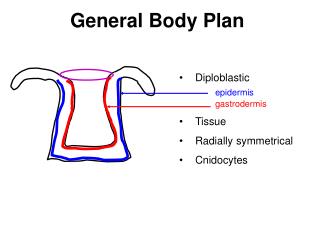
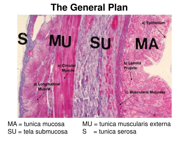
The General Plan. a) Epithelium. b) Lamina Propria. a) Circular Muscle. a) Longitudinal Muscle. c) Muscularis Mucosae. MU = tunica muscularis externa S = tunica serosa. MA = tunica mucosa SU = tela submucosa. Esophagus. tunica mucosa . tela submucosa . tunica

E N D
The General Plan a) Epithelium b) Lamina Propria a) Circular Muscle a) Longitudinal Muscle c) Muscularis Mucosae MU = tunica muscularis externa S = tunica serosa MA = tunica mucosa SU = tela submucosa
Esophagus tunica mucosa tela submucosa tunica musularis exrterna tunica adventitia
Stomach (fundus region) 1. Translucent convoluted areas are gastric pits in tunica mucosa 2. below are “fundic” gastric glands in tunica mucosa
Duodenum Duodenal glands
Small Intestine, Duodenum Pointer is on doudenal (Brunner’s) glands in the tela submucosa the layer (tunica) above is just the general view of the tunica mucosa with intestinal glands
Brunners gland • Small Intestine, Duodenum (100x). • Pointer shows Duodenal (Brunner's) glands in the tela submucosa, their ducts pass through the muscularis mucosa
Small Intestine, Ileum Payer’s patch
Large Intestine, Colon • Pointer - Mucus glandswith many goblet cells. Mucus gland
Liver (lobule) • Central vein of the Liver, (central pointer) • Hepatocytes (everything around it, dark staining) • Sinusoid (capillaries)
The Central Vein (drain) Pointer at Lumen of central Vein
2. 1. Pointer is in the lumen of a branch of hepatic portal vein 2. Pointer is on a branch of the hepatic artery 3. Pointer is on a bile duct (simple cuboidal epithelium) Hepatocytes (cells) = dark staining all around w/ nucleus 3. 1.
The Pancreas Pancreatic islets Pancreatic acini
The Pancreas • Pointers show pancreatic islets • Everything else around are pancreatic acini
Trachea tunica mucosa tela submucosa hyaline cartilage Pseudostratified Ciliated Columnar Epithelium (with goblet cells) at blue pointer.
Trachea 1) pseudostratified ciliated columnar epi2) tracheal cartilage ring3) trachealis muscle 1) 2) 3)
Liver, Portal Area • Branch of hepatic portal vein (Biggest Lumen) • Bile duct (smallest circle, top pointer) • Hepatic artery (thick walled)