Download
1 / 24
250 likes | 570 Vues
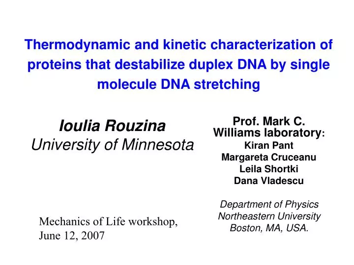
Thermodynamic and kinetic characterization of proteins that destabilize duplex DNA by single molecule DNA stretching. Ioulia Rouzina University of Minnesota. Prof. Mark C. Williams laboratory : Kiran Pant Margareta Cruceanu Leila Shortki Dana Vladescu Department of Physics

E N D
Thermodynamic and kinetic characterization of proteins that destabilize duplex DNA by single molecule DNA stretching Ioulia Rouzina University of Minnesota Prof. Mark C. Williams laboratory: Kiran Pant Margareta Cruceanu Leila Shortki Dana Vladescu Department of Physics Northeastern University Boston, MA, USA. Mechanics of Life workshop, June 12, 2007
Stretching single DNA molecules Microscope objectives Laser beam Polystyrene bead Laser beam DNA molecule F Torsionally relaxed DNA Glass Micropipette
DNA overstretching is a force-induced melting transition Nonequilibrium strand separation dsDNA theoretical Equilibrium melting transition ssDNA G • dsDNA stretch DNA melting free energy G is area under transition region between dsDNA and ssDNA when transition is reversible
HIV-1 NC destabilizes ds DNA and facilitates DNA strand recombination No protein HIV NC Force, pN Extension per base pair, nm “Fast” duplex destabilizers lower melting force and have little hysteresis
Poly Lysine promotes DNA strand annealing more efficiently than wt HIV-1 NC or zinc-less NC, but does not facilitate conversion • DNA stretching: • Increased melting force suggests lack of duplex destabilization; • Very small hysteresis upon relaxation suggests strongly facilitated strand re-annealing
0.2 0.3 0.4 0.5 0.6 0.7 - - - - - + + + + + HTLV-1 NC exhibits stretching profile characteristic of a slow single-stranded binding protein No protein No protein gp32 DNA stretching force , pN HTLV-1NC gp32* DNA extension per bp, nm • Dependence of melting force on pulling rate suggests slow protein binding to ss DNA • The observed large hysteresis indicates that HTLV-1 NC does not easily dissociate single-stranded DNA • Cooperativity of binding to DNA may result in slow dissociation protein negatively charged nucleic acid
SSB proteins are essential for DNA replication Single-stranded DNA binding (SSB) proteins bind to ssDNA and prevent the formation of hairpin structure during DNA replication. T4 Gene 32 dsDNA Fork movement Bacteriophage T4 replication fork http://www.molbio.uoregon.edu/~pvh/images/DNARfork.gif
T4 Gene 32 protein (gp32) • Full length gp32 • Binds preferentially to ssDNA • Highly cooperative binding • Does not lower Tm of dsDNA • Fragment *I • Lacks 48 C-terminal residues • Lowers Tm by ~ 50 °C • Fragment *III • Lacks C- and N-terminal domains • Lacks cooperative binding • Lowers Tm *III 1 21 301 253 - C N- *I Cooperative binding to ssDNA: protein-protein interactions require N-terminus
*I lowers DNA melting force in high salt, gp32 does not No protein stretch No protein relax gp32 stretch gp32 relax *I stretch *I relax 200 nM protein 100 mM Na+ *I retains cooperative binding, lacks C-terminal domain
Measurement of the equilibrium DNA melting force and relaxation kinetics kinetics in the presence of gp32 and *I Force relaxation at constant DNA extension beyound B-DNA length Relaxation time constant Equilibrium melting force
Single molecule measurement of equilibrium binding of gp32 and *I to ssDNA *I gp32 200 mM Na+ 150 mM Na+ 100 mM Na+ 75 mM Na+ 50 mM Na+
Single molecule Kss results for gp32 and *I agree with and extend previous bulk studies
2. Kinetics of individual gp32 binding events Melting transition occurs when: Extension rate = rate of DNA melting due to protein binding no protein 250 nm/s 200 nM *I 25 nm/s gp32 or *I melting melting
Kinetics of reaction from pulling rate dependence of DNA overstretching force • Binding rate is product of • probability of melting n base pairs • ssDNA-protein association rate ka linear approximation measured Apply force:
Mechanical measurement of binding site size and protein association rates only DNA *III 200 nM gp32 400 nM gp32 n=7 ± 1 bp binding site size for gp32 from slope of Fk vs ln(v) 50 nM *I ka from intercept of Fk vs ln(v) with only DNA stretching curve 100 nM *I 200 nM *I
Protein concentration dependence of ka • 3 order of magnitude difference in rates between *I and gp32 • Rates for *I greater than diffusion limit • Stronger than linear dependence of rates on protein concentration 106 100 mM Na+ *I 105 104 Protein binding rate ka (s-1) 3-D diffusion limit 103 102 gp32 101 Protein concentration (nM)
Concentration dependence of ka Fragment *I Full length gp32 50 mM Na+ 75 mM Na+ 100 mM Na+ 150 mM Na+ 200 mM Na+ Kds– binding constant to dsDNA C - protein concentration n - binding site size in bp ks – 1D sliding rate, ~106 s-1 Kds is obtained directly from fits to data McGhee and Von Hippel isotherm
Single molecule measurement of equilibrium binding of gp32 and *I to dsDNA gp32 shows no salt dependence *I shows weaker salt dependence than at higher salt Never previously measured for *I
Core Core Core Core Cationic DNA binding site Unbound *I gp32 bound cooperatively to ssDNA C-terminal domain (CTD) Unbound gp32 N N Core Core Core Na+ Core DNA N N Model for salt-dependent gp32 binding to DNA gp32 binding to ssDNA is regulated by the strongly salt dependent opening of its C terminal domain (CTD) In high Na+ Pop- probability of opening CTD, - free energy of CTD binding to DNA binding site
Another SSB case study: gp2.5 from T7 bacteriophage gp2.5 is a faster SSB then gp32 (less hysteresis; complete re-annealing)
CTD truncation of gp2.5 is faster then wt gp2.5 Equilibrium is reached at ~5nm/s: we can determine protein dissociation rate
Determining Protein Dissociation rate from rate dependence of DNA unwinding C1<C2 F Fm0 Fm1 Fm2 Ln(pulling rate) ka1 ka2 kd
Alternative approach to determine protein dissociation rate by DNA relaxation D CTD gp2.5 Wt gp2.5 Results: dissociation becomes slower in lower salt Dissociation is faster for D CTD mutant of gp2.5
Conclusions • Equilibrium force-induced melting • New method for measuring binding constant to ssDNA • Results match bulk data where available, extend data to new conditions • Rate dependence of overstretching force • New method for measuring protein binding rates • Explains inability of gp32 to lower Tm • 1D diffusion-enhanced kinetics of DNA melting • Rates governed by binding strength, diffusion, and protein conformational changes • Measurements of equilibrium binding constants to dsDNA • gp32 binding regulated by salt-dependent conformational change • Governed by counterion condensation of sodium on DNA and CTD • Electrostatic model explains all data on gp32 binding for first time • CTD binding by replication proteins may also regulate DNA binding • Two complementary approaches to measure protein dissociation rates from ssDNA • By force relaxation to equilibrium at constant extension after fast DNA release - By determining the slow puling rate where DNA stretching curves become rate-independent