Download
1 / 35
390 likes | 983 Vues
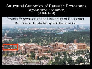
Parasitic Protozoans. Lecture 4. 2- Flagellates. 1- Trypanosoma spp. 2- Leishmania spp. 3- Giardia spp. 4- Trichomonas spp. 1- Trypanosoma spp. 2- Leishmania spp. 1- Hemoflagellates. Called Hemoflagellates because they have a flagellum and require blood medium to culture them.

E N D
Parasitic Protozoans Lecture 4
2- Flagellates • 1- Trypanosoma spp. • 2- Leishmania spp. • 3- Giardia spp. • 4- Trichomonas spp.
1- Trypanosoma spp.2- Leishmania spp. 1- Hemoflagellates • Called Hemoflagellates because they have a flagellum and require blood medium to culture them. • A.K.A. Kinetoplasta • Flagellum is attached to an undulating membrane attached to a kinetoplast. • There is evidence for sexual reproduction but when it occurs is not known. • They can also absorb and use foreign DNA.
1- Hemoflagellates • Two host life cycle • Humans and their domestics are Definitive Host • Insect vectors are the Intermediate Host • Four life stages • Not all stages occur in all species • Certain stages are found in specific hosts • Epimastigotes and promastigotes in insect IH • Amastigoes and trypomastigotes in DH
1- Trypanosoma spp. 1- Trypanosoma gambiensi 2- T. rhodesiensi 3- T. curzi 4- T. lewisi
1- Trypanosoma gambiensi 2- T. rhodesiensi • Definitive Host:Humans. Not pathenogenic to any other species. Native ruminates serve as reservoirs for T.b. rhodesiense, but not T.b. gambiense. • Intermediate Host:Tsetse fly (Glossina) • Mode of transmission: Bite of infected tsetse fly Tsetse fly (Glossina)
Geographic Distribution T. b. gambienseis found in west central and central Africa. T. b. rhodesiensefound in central and east central Africa T. b. gambiense T. b. rhodesiense
1- Trypanosoma gambiensi 2- T. rhodesiensi • Location:Throughout the body in the blood and tissues • Pathology:Both subspecies cause African Sleeping Sickness. • T.b. gambiense causes chronic, long-term form. • T.b. rhodesiense causes an acute form. • Starts with a small sore at bite. • Trypimastigotes divide rapidly and spread throughout body
Pathology • Pathology (con’t): Lymph nodes become swollen and congested • Particularly the nodes in neck • Called Winterbottom’s sign
Pathology • T.b. gambiense frequently goes to CNS • Causes the chronic, sleepiness associated with African Sleeping Sickness • Apathy, mental dullness, disturbance of coordination • Increase in sleepiness, finally to coma, and death. • Death may also occur from malnutrition, falling, or other infections
Pathology • T.b. rhodiensienserarely invade the CNS but causes death much faster. • Usually due to invasion of heart tissue • Both subspecies produce intermittent periods of fever, particularly in early stages. • Due to antigen shifts of the parasite. • They can also take antigens from host body and put them on their body • Much pathology may be due to heightened immune response killing uninfected body cells.
Diagnosis • Trypomastigotes in the blood smear. • Can also be in cerebrospinal fluid • Serological test available
3- Trypanosoma curzi • Definitive Host: Humans, dogs, cats, opossums, armadillos, and wood rats. • Intermediate Host: Reduviid bugs (Kissing bug or assassin bugs). • Location in the Definitive Host:Throughout the body. • Trypomastigotes in blood • Amastigotes most common in spleen, liver, and muscles, including heart • Mode of Transmission: Host rubs tryps into bite wound.
Geographic Distribution Throughout much of central and South America. 12-19 million infected Annual incidence 561,000 2-3 million with chronic symptoms 45,000 die from disease every year. A few cases in U.S. in Maryland, Georgia, Florida, Texas, Arizona, New Mexico, California, Alabama, and Louisiana.
2- Leishmaniaspp. • Leishmania donovani • L. tropica.
Leishmania donovani • Definitive Hosts: Humans. Reservoir includes most mammals • Intermediate Hosts:Phlebotomus sand fly. • Mode of Transmission: Bite of infected Sand Fly • Location in D.H.: Immune system, including spleen, liver, lymph nodes and bone marrow
Geographic Distribution • Probably originated in Old World • Moved to New World with slave trade
Pathology • Causes Visceral Leishmaniasis • A.K.A. Kala-azar, Dum-Dum Fever • Amastigote is engulfed by macrophage. • Macrophage doesn’t kill amastigote. • Neutrophils and eosinophils will kill amastigotes. • Multiplies, breaks out, and each invades another macrophage. • Also destroys macrophages in the spleen, liver, and lymph nodes • Body starts manufacturing macrophages to replace them. • Results in severe wasting and anemia
Pathology Macrophage Infected with amastigotes of Leishmania
Pathology • Early symptoms include malaise, vomiting, low-grade fevers. • Followed with chronic wasting, anemia, enlargement of abdomen due to greatly enlarged spleen and liver. • Death usually follows in 1-2 years if untreated. • Some people recover spontaneously • Related to age and nutrition • Some people who were treated later develop Post-Kala-azar dermal leishmanoid • “Face bumps” • Repeat of treatment usually clears up the bumps.
Diagnosis • Amastigotes in liver tissue, macrophages, spleen, other organs. • IFA, ELISA tests have been developed but can’t tell between L. donovani and L. tropica. • Need to eliminate possibility of typhoid, paratyphoid, malaria, syphilis, tuberculosis, dyssentery, and relapsing fever which cause similar symptoms.
2- Somato-flagellates • 1- Genital flagellates • Trichomonas spp. • 2- Intestinal flagellates • Giardia spp.
Genital flagellates Trichomonas spp. Trichomonas vaginalis • Definitive Hosts: Humans. Reservoir includes most mammals and birds • Intermediate Hosts: Nothing • Mode of Transmission: during sexual inter course by trophozoite
Geographic Distribution • Trichomonas are found in man, monkeys, rodents, fowls, pigeons, doves, termites and slugs. • Distributed in all countries
Pathology • Causes milky yellowish irritant vaginal discharge in female • Ulceral discharge may occur in male
Diagnosis • In female: Examination of vaqginal discharge for trophozoites and urine sample • In male: Examination of prostatic fluid and urine sample
Intestinal flagellates Giardia spp. • Definitive Hosts: Humans. • Intermediate Hosts: Nothing • Mode of Transmission: quadri-nucleated cyst in contaminated food and drink • Flies and cockroaches play an important role in transmission
Geographic Distribution Worldwide, more prevalent in warm climates
Pathology • Children are affected • Mucus production, diarrha, dehydration, intestinal pain, weight loss.
Diagnosis • Microscopic examination of fecal material for identification of trophozoite. • PCR analysis for detection of giardia DNA from both trophozoites and cysts