Download
1 / 57
580 likes | 663 Vues
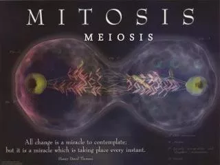
t h e M - p h a s e. M I T O S I S. The process of cell replication. Genes and Proteins. Proteins do the work of the cell: growth, maintenance, response to the environment, reproduction, etc.

E N D
t h e M - p h a s e M I T O S I S The process of cell replication
Genes and Proteins • Proteins do the work of the cell: growth, maintenance, response to the environment, reproduction, etc. • Proteins are chains of amino acids. The sequence of amino acids in each protein is coded in the DNA as a specific sequence of A, C, G and T bases: a gene. • Each gene codes for a different protein.
Genes and Proteins • Key points: • All cells within an organism have the same genes. • What makes cells different from each other is that different genes are turned on and turned off in different cells. • The DNA must be copied and then divided exactly so that each cell gets an identical copy.
MITOSIS VS. MEIOSIS • Mitosis is normal cell division, which goes on throughout life in all parts of the body. Meiosis is the special cell division that creates the sperm and eggs, the gametes. We will discuss meiosis separately. • Mitosis and meiosis occur in eukaryotes. Prokaryotes use a different method—”binary fission” to divide.
NUMBERS OF CHROMOSOMES • Humans have 46 chromosomes • 23 from each parent • Every cell has the same 46 chromosomes • Each species has a characteristic number of chromosomes: • corn has 20, • house flies have 10, • chimpanzees have 48.
SOME VOCABULARY • Chromosomes exist in 2 different states: • Chromatin: Between cell divisions, DNA/protein complex is loosely coiled (easier for protein synthesis) • Chromatid: Right after DNA replication, the chromosomes are tightly coiled together (it is easier to “arrange” chromosomes this way) • There are 2 copies of the chromosome • Centromere: The two copies of the chromatid after replication are held together by the centromere.
CELL CYCLE • Some cells divide constantly: cells in the embryo, skin cells, gut lining cells, etc. Other cells divide rarely or never: only to replace themselves. • Actively dividing cells go through a cycle of events that results in mitosis. Most of the cycle was called “interphase” by the microscopists who first studied cell division. During interphase the cell increases in size, but the chromosomes are invisible. • The 3 stages of interphase are called G1, S, and G2.
INTERPHASE • Interphase is the normal part of cellular function. It includes the following (about 90% of cell life): • G1 phase • S phase • G2 phase
http://www.biology.arizona.edu/cell_bio/tutorials/cell_cycle/cells2.htmlhttp://www.biology.arizona.edu/cell_bio/tutorials/cell_cycle/cells2.html metaphase anaphase telophase prophase M I T O S I S 1 G2 (Gap 2) M cytokinesis 2 G1 (Gap 1) THE CELL CYCLE G0 S-Phase (DNA Self- Replication)
G1 (GAP 1) PHASE • G1 (“Gap”) is the period between mitosis and S, when each chromosome has 1 chromatin (not chromatid). Cells spend most of their time in G1: it is the time when the cell grows and performs its normal function. Control of cell division occurs in G1: a cell that isn’t destined to divide stays in G1, while a cell that is to divide enters the S phase.
S PHASE • The S phase (“Synthesis”) is the time when the DNA is replicated, when the chromosome goes from having one chromatin to having 2 chromatids held together at the centromere.
G2 (GAP 2) PHASE • G2 is the period between S and mitosis. The chromosome have 2 chromatids, and the cell is getting ready to divide.
HOW TO IDENTIFY INTERPHASE Pictures of cells during interphase
nucleolus (if any) still visible Interphase nuclear envelope clearly visible chromatin, NO chromosomes, yet http://www.fed.cuhk.edu.hk/~johnson/photomicrographs/mitosis/animal/animal_interphase.htm
INTERPHASE http://iccbweb.med.harvard.edu/mitchisonlab/Pages/mt.html
interphase interphase
INTERPHASE Allium root tip Coregonus blastula
INTERPHASE is the normal lifetime of a cell, after being “born” by division, and before it divides itself. MITOSIS INTERPHASE is not a stage of mitosis ! Biological Science, a Molecular Approach. BSCS Blue Version. Heath and Company, 1996.
What is MITOSIS ? THE PROCESS BY WHICH TWO NEW NUCLEII ARE FORMED, WITH EXACTLY THE SAME KIND AND NUMBER OF CHROMOSOMES AS THE PARENT CELL. (1 CELL TO 2 CELLS) http://fairmanstudios.com/als.htm
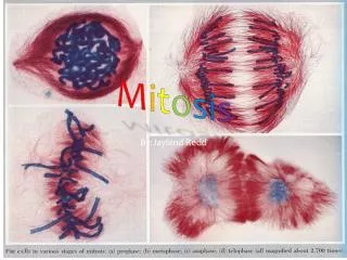
STEPS IN MITOSIS (In AP, the phases are not emphasized) • PROPHASE • METAPHASE • ANAPHASE • TELOPHASE
PROPHASE • In prophase, the cell begins the process of division. • The chromosomes condense. The proteins attached to the DNA cause the chromosomes to go from long thin structures to short fat one, which makes them easier to pull apart (VISIBLE). • The nuclear envelope disappears. The double membrane that surround the nucleus dissolves into a collection of small vesicles, freeing the chromosomes to use the whole cell for division • Centrosomes form and move to opposite poles. During interphase, the pair of centrosomes were together just outside the nucleus. In prophase they separate and move to opposite ends of the cell. • The spindle starts to form, growing out of the centrosomes towards the chromosomes.
PROMETAPHASE • Nuclear membrane fragments • Spindle interaction with chromosomes • Kinetochore develops at the centromere (this is where the spindle microtubules are going to bind)
HOW TO IDENTIFY PROPHASE Pictures of cells during prophase
PROPHASE • Nuclear membrane dissolves. • Nucleolus disappears. • Chromosomes form. • Centrioles migrate & form spindle. http://www.blc.arizona.edu/courses/181gh/Lectures_WJG.01/mitosis_F.01/mitosis.html
PROPHASE nuclear envelope disappears nucleolus disappears chromosomes become visible http://www.ac-dijon.fr/pedago/svt/documents/mitose/prophase.gif
PROPHASE http://www.itg.uiuc.edu/technology/atlas/structures/mitosis/prophase.htm
PROPHASE Allium root tip Coregonus blastula
METAPHASE • Metaphase is a short resting period where the chromosomes are lined up on the equator of the cell, with the centrosomes at opposite ends and the spindle fibers attached to the kinetochore. Everything is aligned for the rest of the division process to occur.
HOW TO IDENTIFY METAPHASE Pictures of cells during metaphase
METAPHASE chromatids spindle centriole http://www.chembio.uoguelph.ca/educmat/chm736/cycletx.htm • chromatids line up on the equator.
METAPHASE TWO IDENTICAL COPIES OF ONE CHROMOSOME. THIS CHROMATID WILL SOON MOVE TO NORTH POLE THIS CHROMATID WILL SOON MOVE TO SOUTH POLE http://genenlab.spoono.com/gnu/mandm.shtml
chromatids spindle centriole Nature (408. 423, 2000). http://www.blc.arizona.edu/courses/181gh/Lectures_WJG.01/mitosis_F.01/mitosis.html
METAPHASE http://iccbweb.med.harvard.edu/mitchisonlab/Pages/mt.html
METAPHASE Allium root tip Coregonus blastula
ANAPHASE • In anaphase, the centromeres divide. At this point, each individual chromosome goes from: • 1 chromosome with 2 chromatids to: • 2 chromosomes with one chromatid each. • Then the spindle fibers contract, and the chromosomes are pulled to opposite poles, towards the centrosomes.
HOW TO IDENTIFY ANAPHASE Pictures of cells during ANAPHASE
ANAPHASE • chromatids migrate to each pole. http://www.blc.arizona.edu/courses/181gh/Lectures_WJG.01/mitosis_F.01/mitosis.html
ANAPHASE http://www.univ-orleans.fr/SCIENCES/BIOCHIMIE/MMC/accueil.htm
ANAPHASE early late Conly Rieder http://www.wadsworth.org/BMS/SCBlinks/WEB_MIT2/HOME.HTM
ANAPHASE Allium root tip Coregonus blastula
TELOPHASE • In telophase the cell actually divides. • The chromosomes are at the poles of the spindle. • The spindle disintegrates • The nuclear envelope re-forms around the two sets of chromosomes (become less coiled). • The cytoplasm is divided into 2 separate cells, the process of cytokinesis.
CYTOKINESIS • The organelles (other than the chromosomes) get divided up into the 2 daughter cells passively: they go with whichever cell they find themselves in. • Plant and animal cells divide the cytoplasm in different ways. • In plant cells, a new cell wall (CELL PLATE)made of cellulose forms between the 2 new nuclei, about where the chromosomes lined up in metaphase. Cell membranes form along the surfaces of this wall. When the new wall joins with the existing side wall, the 2 cells have become separate. • In animal cells, a ring of actin fibers (microfilaments are composed of actin) forms around the cell equator and contacts, pinching the cell in half. (CLEAVAGE FURROW)
CELL CYCLE CONTROL • These will control whether a cell divides or not • Growth Factor: a protein that is released that induces a cell to divide (cell communication) • Density-Dependent Inhibition: if an area is too crowded with cells, cell division is inhibited. If the area lacks cells, division is allowed to occur • Anchorage Dependence: must be attached to a substrate
HOW TO IDENTIFY TELOPHASE Pictures of cells during ANAPHASE
TELOPHASE http://www.bmb.psu.edu/courses/bisci2/mitosis/mitosis.htm • Chromosomes dissolve. • Mitotic spindle dissolves. • Nuclear membrane forms. • New nucleoli form.
TELOPHASE ONE DAUGHTER NUCLEUS FORMS AT NORTH POLE ONE DAUGHTER NUCLEUS FORMS AT SOUTH POLE SPINDLE APPARATUS DISSOLVES
TELOPHASE early late New nuclei form at the poles. Cytokinesis begins. http://www.blc.arizona.edu/courses/181gh/Lectures_WJG.01/mitosis_F.01/mitosis.html
TELOPHASE Allium root tip Coregonus blastula
Summary of Mitosis • Prophase: • Chromosomes condense • Nuclear envelope disappears • centrosomes move to opposite sides of the cell • Spindle forms and attaches to centromeres on the chromosomes • Metaphase • Chromosomes lined up on equator of spindle • centrosomes at opposite ends of cell • Anaphase • Centromeres divide: each 2-chromatid chromosome becomes two 1-chromatid chromosomes • Chromosomes pulled to opposite poles by the spindle • Telophase • Chromosomes de-condense • Nuclear envelope reappears • Cytokinesis: the cytoplasm is divided into 2 cells