Download
1 / 26
260 likes | 265 Vues
Join us at the 7th East African Health and Scientific Conference in Tanzania to learn about the latest advancements in Liquid Chromatography-Mass Spectrometry (LC-MS) techniques in drug discovery. Discover how LC-MS is revolutionizing the field and providing invaluable insights into drug analysis. Don't miss out on this informative event!

E N D
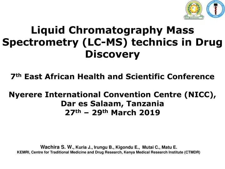
Liquid Chromatography Mass Spectrometry (LC-MS) technics in Drug Discovery 7th East African Health and Scientific Conference Nyerere International Convention Centre (NICC), Dar es Salaam, Tanzania 27th – 29th March 2019 Wachira S. W., Kuria J., Irungu B., Kigondu E., Mutai C., Matu E. KEMRI, Centre for Traditional Medicine and Drug Research, Kenya Medical Research Institute (CTMDR)
Background • Mass spectrometry (MS) was born in 1897, by Sir J. J. Thomson he was awarded the Nobel Prize in Physics in 1906. • At the beginning of the 20th century he constructed the first mass spectrometer, sorting ions according to their trajectory in the electromagnetic field. • Thomson’s scholar F. W. Aston (1922 Nobel Prize in Chemistry) and A. J. Dempster improved resolution and developed the first ionisation technique EI (electron ionisation). • Chemists sought high resolution mass analyzers, being able to detect elements and small molecules, which led to the development of time-of-flight and quadrupole mass spectrometers.
Background • Those could be coupled to gas chromatography (GC) and later in the 1950s to liquid chromatography (LC) by W. Paul, who was awarded the Nobel Prize in Physics in 1989 for his evolutions in ion-trap MS. • coupling to liquid chromatography were achieved, in the 1980s still no large molecules could be analyzed as these were fragmented already during ionisation or evaporation. • It was only when J. B. Fenn applied the soft ionisation technique ESI (electrospray ionisation) to peptides and F. Hillenkamp, M. Karas and K. Tanaka developed the MALDI technique (matrix assisted laser desorption ionisation), biological experiments with biological macromolecules became possible. • J. B. Fenn and K. Tanaka were awarded with part of the Nobel Prize in Chemistry in 2002 for this work
Why Liquid Chromatography • Analysis of labile analytes • Analysis of more polar compounds without derivatization. • Analysis of significantly higher masses • Reduction of lengthy clean-up using HPLC
Mass spectroscopy • MS is the most versatile and comprehensive • It measures the masses of individual molecules, fragments of molecules and atoms. • It provides ultra high detection sensitivity • The mass spectrum of each compound is unique and can be used as chemical “fingerprint” together with its retention time to characterize the compound.
Liquid Chromatography Coupled With Mass Spectroscopy • LC with detectors like Refractive index, electrochemical, fluorescence, and ultraviolet-visible (UV-Vis) detectors generate two dimensional data; that is, data representing signal strength as a function of time • When coupled with MS In addition to signal strength, they generate mass spectral data that can provide valuable information about the molecular weight, structure, identity, quantity, and purity of a sample. • LC/MS is a hyphenated technique combining the separation power of HPLC, with the detection power of mass spectrometry
Liquid Chromatography Coupled With Mass Spectroscopy • It uses an interface that will eliminate the solvent and generate gas phase ions, transferred to the optics of the mass spectrometry • Obtain spectra and molecular mass identification for each peak eluted from the chromatography column. • Straightforward mass spectra of directly infused samples won't distinguish between, buffer components, contaminants and other components of a sample mixture.
Instrumentation & chromatographic conditions • The LC system equipped with binary pump, an autosampler set at 15°C, an online solvent degasser and column oven is used to inject 1-10 μl aliquots of the processed samples on a C18 analytical column • Separation and elution is achieved by gradient system using solvent (formic acid, methanol, water) as the mobile phase, at a flow rate that is determined • The total LC run time is determined • LC–MS detector analysis is performed with IonSpray source interface in positive ionization mode. • Direct infusion of 10-100 ng/ml of each analyte in methanol is made by syringe pump to optimize parameters to detect the most vivid signals of transitions from precursor to product ions.
LC-MS/MS method development • Bioanalytical LC-MS/MS method development comprises three main parts: • First, MS/MS spectra are recorded and mass spectrometry settings are optimized, to find a specific fragment ion and the most appropriate system parameters. • Second, a fast liquid chromatography method, suitable for MS/MS detection, is developed to separate the analytes from matrix components. • Third, a cost-effective and selective sample preparation technique is selected and optimized, supporting the analysis by LC-MS/MS.
LC-MS/MS method development • Molecular ions, produced in the ion source at atmospheric pressure (760 torr, 1013 mbar), enter the first region of the mass spectrometer via a small orifice; here through a so-called “curtain gas” (inert gas), which assists declustering from solvent molecules. • This part of the mass spectrometer is evacuated by a first turbo pump and a backing pump to ensure a high vacuum and avoid collisions with other molecules. • In this region, the molecular beam is focused and led through a first quadrupole, the Q0. • Focussing in this first region is achieved in different ways in different mass spectrometers.
LC-MS/MS method development • The actual mass analysis takes place in the high vacuum region (10-5 torr), which is evacuated by a second turbo pump and backing pump. • Additional stabilizers and lenses are involved to keep the ions on their path towards the detector, which is in this case a channel electron multiplier. • Two main principles accelerate the ions into and through the mass spectrometer: the vacuum gradient and the DC voltage differential • Quadrupole mass spectrometers are also called mass filters, as they have the ability to selectively filter ions with a given mass-to-charge ratio (m/z) by a combination of direct current (DC) and alternating current (AC), applied to each quadrupole.
Method validation • The method validation is very important especially in in vivo clinical trials • It is done some days before the commencement of the analysis • This is done in accordance with the existing guidelines for specificity, selectivity, sensitivity, linearity, precision, accuracy, recovery and Stability
Isolation and identification of compounds from R. communis • The methanol extract of R. communis was subjected to vacuum liquid chromatography using silica gel eluting with hexane, ethyl acetate, methanol and water. • Three fractions were obtained. Fraction 2 was subjected to repeated column chromatography eluting with ethyl acetate, methanol and water and this yielded two pure compounds identified by LC-MS as • Ricinine and • 3-Carboxy-4-methoxy-N-methyl-2-pyridone.
Identification of compounds from R. communis 3-Carboxy-4-methoxy-N-methyl-2-pyridone Conversion of Ricinine to its carboxylic acid
Identification of compounds from R. communis 3-Carboxy-4-methoxy-N-methyl-2-pyridone
Uses of Mass Spectrometryin Drug Discovery • Qualitative Analysis • Elucidation of the structural characteristics of various substances in different matrices: • What is in the sample? • Quantitative Analysis • Determination of the concentration of various substances in different matrices: • How much is in the sample?
Other Uses • Useful for analyzing large biomolecules such as proteins, peptides,and oligonucleotides • Analyze smaller molecules like benzodiazepines,sulfated conjugates • Electrospray can be used to analyze molecules as large as 150,000 u even though the mass-to-charge range for a typical LC/MS instruments is around 3000 m/z.
Comparison to other methods • This bio-analytical method has several advantages • Sample preparation is simpler and the chromatographic column and IS used are commercially available. • The procedure for sample preparation and extraction are rapid and inexpensive • The method is very sensitive and selective • Another advantage of this method is the use of a gradient mobile phase of simple composition with reduction in run time of analysis, which allows determination of the analytes more precisely. • It has a wide range of use