Download
1 / 17
170 likes | 354 Vues
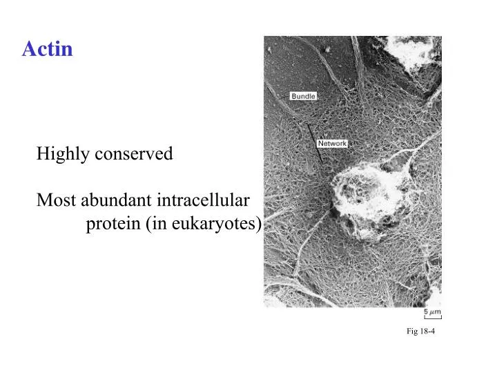
Actin. Highly conserved Most abundant intracellular protein (in eukaryotes). Fig 18-4. Structure of actin. Polarity of molecule ‘-” and ‘+’ ends T form and D form G- actin vs F- actin. Fig 18-2. Structure of actin. View from ‘-’end. Fig 18-2. Bundles and Networks.

E N D
Actin Highly conserved Most abundant intracellular protein (in eukaryotes) Fig 18-4
Structure of actin Polarity of molecule ‘-” and ‘+’ ends T form and D form G- actin vs F- actin Fig 18-2
Structure of actin View from ‘-’end Fig 18-2
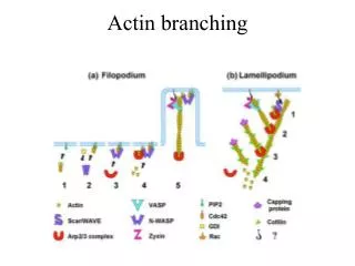
Bundles and Networks What is the benefit of these kinds of associations?
Stability of actin Stability depends on environment– ion and g-actin concentration Provides support through DYNAMIC arrangements including both structure and gel-like qualities of cytosol
Actin filaments dynamics Filaments utilizes 3 steps: lag period, elongation, steady state ATP hydrolysis NOT required for polymerization ATP hydrolysis changes kinetics of polymerization + vs - end? http://www.bms.ed.ac.uk/research/smaciver/lectures/Cs2.htm Illustration of treadmilling CBI 25.2
Microtubules Protofilament: Polymer of a /b tubulin heterodimers 13 protofilaments= microtubule Stable and unstable populations Exhibit dynamic instability Fig 19-5 Fig 19-1
Microtubule dynamics http://www.bms.ed.ac.uk/research/smaciver/lectures/Cs2.htm + vs - end Steps in formation protofilament formation microtubule assembly microtubule elongation Microtubules treadmill AND undergo dynamic instability
Factors effecting polymerization/ depolymerization Critical Concentration: Cc Actin: ends have different Cc (+) 0.1mM (-) 0.8mM cellular concentration 0.5mM Consequences? Microtubules: One end ‘in’ MTOC Consequences? Associated proteins Toxins
And why are we discussing this? Shmoos form from reorganization of actin cytoskeleton Shmoo tip ‘extends’ due to vesicles specifically delivered via actin bound motor protein Microtubules are main ‘highway’ vesicular traffic
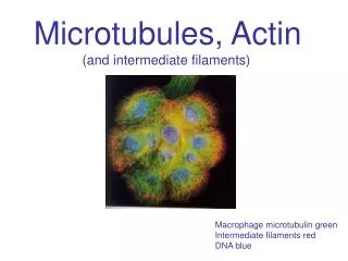
Why important for neuronal function? Fig 23-30 Actin in development CBI21.5 Microtubules in axon core and protein localization Intermediate filaments in mechanical stability of neurons
What makes one MT different from another? Accessories!
Toxins Effecting Actin Cytocholasin D Latrunculin Phalloidin Bind + ends Bind G-actin Bind between subunits on F-actin Effecting Microtubules Colchicine Colcemid Taxol Vinblastine Block formation by binding MT + end (favors depolymerization) Same as colchicine but reversible Stabilizes by binding sides Lo concentrations act like taxol: hi promotes crystalline arrays Net effect on cell-- death Research and Medicinal purposes for some
Actin associated Microtubule associated Associated proteins: cellular Roles of associated proteins
Could this relate to Bipolar Disorder? http://cellbio.utmb.edu/cellbio/microtubule_structure.htm#MAPs
Next time Introduction to molecular motors