Download
1 / 1
10 likes | 76 Vues
Rescue Mutation T381A. 70. 50. 47. 60. 45. 37.6. 50. 40. 34.1. 40. 35. 33.7. 30. 30. 25. 20. 20. 10. 12. 11.76. 14. 15. 0. 1. 55. 28. 37. 46. 73. 91. 10. 19. 64. 82. 100. 109. 118. 127. 163. 136. 145. 154. 10. 5.23. No Pro. TP. P. 5. 1.17. 0. 0.

E N D
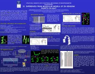
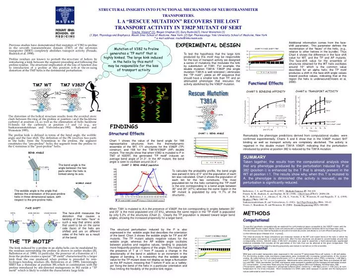
Rescue Mutation T381A 70 50 47 60 45 37.6 50 40 34.1 40 35 33.7 30 30 25 20 20 10 12 11.76 14 15 0 1 55 28 37 46 73 91 10 19 64 82 100 109 118 127 163 136 145 154 10 5.23 No Pro TP P 5 1.17 0 0 0 0 0 0.58 0 0 0 0 0 0 0-5 5-10 10-15 15-20 20-25 25-30 30-35 35-40 40-45 CHART 3:WOBBLE ANGLE CHART 4: FACE-SHIFT TM7 TP motif (V382P mutant) Proline in T381A V382P mutant 200 150 150 100 100 50 50 0 0 -50 -50 - 100 -100 -150 -150 -200 TP P No proline TP P 12 Wild type 10 T381A 8 H]-serotonin uptake (pmol/mg protein) T381V 6 4 2 T381A-V382P [ 3 0 T381V-V382P, V382P 0 10 20 30 40 50 Time (min.) STRUCTURAL INSIGHTS INTO FUNCTIONAL MECHANISMS OF NEUROTRANSMITER TRANSPORTERS. I. A “RESCUE MUTATION” RECOVERS THE LOST TRANSPORT ACTIVITY IN T382P MUTANT OF SERT Irache. Visiers* (1), Megan Stephan (2), Gary Rudnick(2), Harel Weinstein (1) (1 )Dpt. Physiology and Biophysics. Mount Sinai School of Medicine, New York. (2) Dpt. Pharmacology. Yale University School of Medicine, New York * e-mail address; irache@inka.mssm.edu Mutation of V382 to Proline generates a ‘TP motif’ that is highly kinked. The large kink induced in the helix by this motif may be responsible for the loss of transport activity Additional information comes from the face-shift parameter,. This parameter defines the reorientation of the “faces” of the helix, (e.g., relative to other helices in the bundle). Thus Chart 4 shows the difference in the face shift value for WT, and the TP and AP mutants. The face-shift value for the ensemble of structures obtained for the WT helix oscillates around 10 which is the common value described for an alpha helix; the TP motif produces a shift in the face-shift angle values toward positive values, indicating that at this region the helix is underwound(Visiers et al, 2000) EXPERIMENTAL DESIGN To test the hypothesis that the large kink produced by this motif may be responsible for the loss of transport activity we designed a series of mutations that modulate the kink by substitution of T381. For example, the double mutation T381A/ T382P (the single mutation T381A is well tolerated ) eliminates the “TP motif”, yields an AP sequencethat should have a smaller kink than TP, and an attenuated phenotype that rescuess the activity abolished by the V382P mutation. Functional Effects TM7 WT TM7 V382P CHART 6: TRANSPORT ACTIVITY CHART 5: BINDING AFFINITY 382 FINDINGS Structural Effects CHART 1: BEND ANGLE Remarkably the phenotype predictions derived from computational studies were confirmed experimentally. Charts 5 and 6 show that in the V382P mutant 5HT binding as well as transport activity are completely abolished. The activity is regained in the double mutant T381A V382P, indicating that the perturbation introduced by proline at position 382 is reduced by the T381A mutation. Chart 1 shows the value of the bend angle for 166 representative structures from the thermodynamic ensemble of the WT, 171 structures for the V382P (TP) construct, and 168 for the T381A/V382P (AP) double mutant. The results show that when V382 is mutated to P in TM7 of hSERT, the generated ‘TP motif’ induces an average bend angle of 31.3. In the AP mutant, the bend angle is seen to oscillate around 24.4. SUMMARY Taken together, the results from the computational analysis show that any phenotype produced by the perturbation induced by P at 382 (position i) is enhanced by the T that is already present in the WT at position i-1. The results show why, when this T is mutated to A, the phenotype is diminished (the activity is rescued), as the perturbation is significantly reduced. The bend angle is the angle between the two parts when the helix is kinked along its axis. CHART 2: BEND ANGLE population To calculate the probability profile, the bend angle was parsed in bins of 5 and the population of each bin was calculated. Chart 2 shows the population of each bin for the two constructs. The most populated bin for the helix containing the ‘TP motif’ is the one corresponding to a bend angle between 30 and 35 (47%) whereas the same region in the AP mutant is populated by only 11.7% of the structures . The wobble angle is the angle that defines the orientation of the post-proline helix in three dimensional space, with respect to the pre-proline helix. When T381 is mutated to A in the presence of V382P, the bin corresponding to angles between 20 and 25 is the most populated one (37.6%), whereas the same region in the ‘TP motif’ is populated by only 5.2% of the structures (Chart 2). Clearly the TP population is skewed toward larger bend angles, showing the increased propensity for a larger bend. METHODS The face-shift measures the distortion that causes a twisting of the helix “face” in such a way that amino acids that used to be on the same side (face) of the helix are shifted and are on different sides of the helix as a result of the bend. Computational To test this hypothesis computationally we explored the conformational space accessible to the V382P mutant and the T381A/V382P double mutant. Monte Carlo simulations with a Scaled Collective Variables (SCV) technique was used. This technicque mimics thermal fluctuations around minimized structures and allows to a more efficient sampling of the conformational space (Hassan et al. in press). The structural perturbation induced by the P is also expressed in the wobble angle that describes the orientation of the bend. Chart 3 shows the wobble angle for TP and AP mutants. The TP mutant adopts negative values for the wobble angle, whereas the AP wobble angle oscillates between positive and negative values, tending to populate more frequently positive regions of the angle. This means that the presence of T at i-1 from P causes a change in the directionality of the bend in addition to an increment in the degree of bending. It is noteworthy that the wobble angle value for the TP mutant does not display as large a fluctuation as the AP mutant, meaning that T before P acts as a ‘safety-pin’ fixing the bend of the helix in a particular orientation and thus limiting the flexibility of the proline kink region. The initial structures for these simulations are molecular models of the WT and mutant forms of the helix in which the dihedral angles correspond to a regular proline kink. After the two structures are allowed to accommodate around their corresponding minima, 500,000 steps of MC-SCV simulation are used to assemble a thermodynamically equilibrated statistical ensemble that includes all the geometries of the helix that can be attained at the given temperature. The probability analysis that yields the bending propensity (see below) is carried out on this ensemble. THE “TP MOTIF” Experimental Wild type and mutant transporters were expressed in the vaccinia-T7 polymerase-HeLa cell system, in 48 well plates. For the binding studies crude membrane preparations were incubated with increasing concentrations of the cocaine analog, 2b-carbomethoxy-3b-(4-iodophenyl)tropane (CIT) in phosphate-buffered saline (PBS) containing 1 mM MgCl2 and 0.1 mM CaCl2, for 1.5 h at room temperature. Membranes were collected on glass fiber filters and washed 3X with ice cold PBS. Mock transfected (no DNA) membranes were assayed in parallel and the background subtracted at each CIT concentration. Curves were fitted using the Origin plotting program (Microcal Software, Northhampton, MA). [3H]-serotonin uptake was measured in phosphate-buffered saline containing 1 mM MgCl2and 0.1 mM CaCl2 at room temperature for the times indicated. Mock transfected (no DNA) wells were assayed in parallel and the background values subtracted at each time point.