Download
1 / 85
920 likes | 1.22k Vues
The Gastrointestinal Tract. Basic Principles. One function of the G.I. tract is to absorb nutrients from the diet. Digestive system can be thought of as a 1-way “tube” for the absorption of nutrients. The G.I. tract must also act as a barrier against ingested toxins. Basic Principles.

E N D
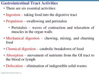
Basic Principles • One function of the G.I. tract is to absorb nutrients from the diet. • Digestive system can be thought of as a 1-way “tube” for the absorption of nutrients. • The G.I. tract must also act as a barrier against ingested toxins.
Basic Principles • Both absorptive and barrier functions of the intestinal tract rely on a monolayer of specialized epithelial cells. • 1-cell layer separates ingested contents from the circulatory system (ie. the rest of your internal organs) • Similar to the respiratory system • Epithelium is polarized (directional) • Apical = facing lumen / food (exterior) • Basal / basolateral = facing circulating blood supply (interior) • Epithelial cells “adhere” to one another very tight • Prevents free passage of solutes from the lumen of the gut into the blood (the only way to get into the blood is THROUGH the cells, not between them)
Unique Features • Enteric nervous system (“brain” in the gut) • G.I. tract interacts with the CNS primarily through the vagus nerve. • Enteric nervous system represents a “stand-alone” neural control network. • Composed of 2 sheets of interconnected neural networks: • Submucosal plexus (below mucosal layer) • Myenteric plexus (between smooth muscle layers) • My-enteric = myo (muscle) enteric (gut) • If you counted up all the neurons in your body, almost ½ are in your digestive system!
Enteric Nervous System Submucosal plexus Myenteric plexus
Unique Features • Enteric motor neurons serve to “relax” intestinal smooth muscle • Without enteric motor neural stimulation, intestinal smooth muscle remains contracted • Hirchsprung’s disease (congenital megacolon) = fetal error in the migration of the enteric nervous system • During fetal development, the gut tube forms from the stomach – rectum • The enteric nervous system must follow …sometimes it stops short • The region of the gut that doesn’t contain any enteric nerves = clamped shut…nothing can pass = “megacolon”
Peristalsis Contraction of circular muscle behind the bolus Contraction of longitudinal muscle IN FRONT of the bolus Peristalsis: coordinated contraction of both circular and longitudinal muscle layers of the G.I. musculature as controlled by the enteric nervous system
Peristalsis There are different forms of peristalsis: segmentation & directional peristalsis. Despite the end-result of one-way directional peristalsis, segmentations occur much more often than directional contractions. Segmentation allows to gut to break up the chyme more effectively.
Unique Features • Remember!!!! • The enteric nervous system cannot sense pain • Pain sense requires “Substance P” receptor • While the intestine can secrete Substance P, the gut does not express any substance P receptors • This makes if quite difficult to diagnose intestinal “cramps” etc. • The pain these patients complain of is somatic (resulting from intestinal derived Substance P acting on the central nervous system)
Unique Features • G.I. tract has a resident mucosal immune system to aid in barrier function. • contains almost 1/2 of the body’s immune cells. • G.I. tract is under constant assault from ingested toxins.
Unique Features • The mucosal immune system: “the enemy of your enemy”. • Alterations can lead to severe pathophysiological responses: • Food allergies • Inflammatory bowel disease, Crohn’s, colitis • Hypoxia-induced necrosis, ischemia-reperfusion injury • Necrotizing enterocolitis
Unique Features • Epithelia of the G.I. tract has a very high turnover/production rate • Surface epithelia will renew itself every 2-6 days depending on the region of the intestinal tract • A very large amount of stem cells in the gut helps to replace the epithelium quickly • Anti-cancer therapies (targeting rapidly dividing cells) often have the side-effect of targeting the gut as well
Epithelial Turnover Stomach Gastric pit/crypt Stem cells at the bottom of the crypt/pit.
Epithelial Turnover Small intestine Villus/Surface Mature enterocytes that absorb nutrients Crypt Stem cells that are immature and secrete water and ions very well
Epithelial Turnover Colonic crypt Stem cells at the bottom of the crypt.
Different epithelial “shapes” Esophagus Stomach Small intestine Large intestine
Cephalic phase • Anticipation of a meal leads to preparation by the stomach • Small amount of acid secretion to “prime” the gastric lumen for ingested bolus • Voluntary: can be triggered by thought • Largely due to CNS influencing the enteric nervous system
Mastication (chewing) • Break up bolus • Mix with saliva • mucus provides lubrication • salivary amylase begins starch breakdown
Swallowing • Voluntarily force the bolus to the back of your mouth (pharynx) with back you your tongue • Remember the respiratory system? The pharynx is the upper region, from the nasal cavity - larynx • Once in pharynx, enteric neural control becomes automatic (involuntary). • Pharynx contracts to push bolus into esophagus • Epiglottis flips down to close off trachea • Upper and lower esophageal sphincters open in unison.
1 2 3
Esophagus The epithelium of the esophagus is unique in the digestive system because it is stratified (many cells thick). Also, the upper region of the esophagus is lined with skeletal muscle and is under voluntary control.
Stomach “processing” • Bolus passes through the lower esophageal sphincter, and enters the stomach • Gastric functions include: • Storage of meal contents • Acid secretion for protein hydrolysis and antimicrobial action • Mixing meal contents into pulp/liquid consistency (chyme)
Lower esophageal sphincter Fundus (storage) 3 muscle layers longitudinal circular transverse Body/corpus (acid & pepsin secretion, storage) Pylorus (resistance) Antrum (acid control & mixing)
Mucus secretion (acid protection) Acid control/acid & pepsin secretion
Stomach “processing” • Acid secretion combined with strong contractions serve to mash the bolus into a liquid consistency. • Pylorus is constantly contracted (never really relaxes/opens) • Only as a liquid (chyme) can the meal be pushed through the pylorus and into the duodenum (small intestine).
Small Intestine (absorption) • Small intestine is the site of majority of nutrient absorption • 3 “regions” = duodenum, jejunum & ileum • Duodenum has numerous sensory cells to detect caloric, pH and fat content of the bolus. • Feedback control over the rate of gastric emptying into the duodenum • Carbohydrates & proteins are permitted to enter faster/sooner than fats • Heavy meals tend to remain in the stomach longer because fats are very calorie “dense” (per gram, fat has the highest amount of calories) • Effort to regulate the rate of calories that enters the small intestine
Small Intestine • Pancreatic digestive enzymes and liver bile both enter duodenum via Sphincter of Oddi • Proteases, amylase (to digest protein and carbohydrates), bicarbonate (buffer pH from stomach acid) from the exocrine pancreas • Bile acids (to control cholesterol levels and emulsify fats for absorption) from the liver • Chyme is gradually propelled down the small intestine (duodenum-jejunum-ileum) • Remember that there are more segmental contractions, even though net-result is one-way propulsion
Small Intestine • Bile acids/bile salts contain bilirubin (exhausted heme) & cholesterol • In patients with gall bladder or liver disease, jaundice can often be accompanied by discolored fecal matter (looks more white/clear) • Result of inadequate bilirubin excretion • If patient cannot transfer bile salts/acids into the fecal stream (into the gut), the body cannot rid itself of these compounds (bilirubin & cholesterol) • If they build up in the blood, they become toxic
Small Intestine • The surface area of the small intestine is 100-200 m2 ( tennis court). • Large surface area provides greater contact for nutrient absorption. • Epithelial surface area is amplified by a number of structures: • Plica (2-5X increase in surface area) • Folds in the intestinal musculature • Villi (10-30X increase in surface area) • Arrangements of the intestinal epithelium (monolayer) • Microvilli of each absorptive enterocyte (200-400X increase in surface area). • Specialized arrangement of the apical plasma membrane
Microvilli 0 minutes 5 minutes Microvillus height can be changed rapidly in response to various stimuli
Oxygen Countercurrent Exchange • Oxygen countercurrent exchange during splanchnic blood evacuation (severe blood loss, low blood flow resulting from C.V. disease) can deprive villus/surface enterocytes of oxygen • Any time there is low blood flow • Oxygen deprivation rapidly leads to enterocyte death. • In extreme cases, massive enterocyte death can lead to necrosis of the intestinal surface, which compromises the barrier function of the intestine
Small Intestine • Nutrients (carbohydrates, proteins) are digested down to their basic building blocks by enzymes: • Monomeric sugars, amino acids, small peptides • Consequently, the absorptive epithelia need only express a few specialized transport proteins in order to absorb nutrients • If you absorbed intact proteins & carbohydrates, you’d need specific transport proteins for each protein and carbohydrate…and there are millions of them • If you break down proteins into the 20 amino acids, you need only 20 amino acid transport proteins (actually, there are about 8) • If you break down carbohydrates into glucose, galactose & fructose (the monomers of carbohydrate), you need 2 “sugar transporters”
Sugar Transport Glucose Glucose Glucose Glucose Na+ Na+ Na+ Na+ Carbohydrates are digested by pancreatic enzymes and enzymes on the microvillus into MONOMERS (glucose). These sugar monomers are then transported into the enterocyte using the sodium gradient. Remember that all cells try to push out sodium and keep very little sodium INSIDE themselves.
Sugar Transport Glucose SGLT-1 Na+ Na+/K+/ATPase SGLT-1 (sodium glucose cotransporter) - secondary active transporter On the lumen-side of the enterocyte is a “secondary active transporter” called the “sodium glucose co-transporter” (SGLT-1). This transport protein first binds sodium. The sodium then changes the shape of this protein, allowing the glucose-pore to open. Once this transporter finds BOTH sodium and glucose, it will change it’s shape and bring BOTH into the cell. The sodium will be pumped out into the blood using the Na/K/ATPase enzyme.
Sugar Transport SGLT-1 Glucose SGLT-1 (sodium glucose cotransporter) - secondary active transporter The glucose will then transfer out of the cell into the blood through another transporter known as GLUT-2. This sugar transporter is different from SGLT-1 because it only relies on a concentration gradient (it is a glucose channel that allows glucose to pass across the plasma membrane according to the concentration gradient. What would happen if you used facilitative transporters (glucose channels) on both sides of the absorptive cell?
Small Intestine • Nutrient absorption is coupled to water absorption. • Water is “dragged” by nutrients to maintain osmolarity • If you didn’t do this, water would remain inside the gut tube, and you would only put electrolytes and nutrients into the blood…what would happen to your blood “tonicity”? • Remember that “ball” of water molecules that surrounds a “dissolved” particle? • Remember the aquaporin molecule that allows water molecules to pass across the plasma membrane? • What is the term for water movement (it’s different from the movement of particles/solutes)? • Nutrient absorption is generally complete by the ileum. • Remaining chyme contains “resistant” nutrients (relevant in the large intestine), and electrolytes.
Small Intestine • Carbohydrates (sugars) and protein (peptides and amino acids) are absorbed into the venous blood and transported into the hepatic portal system destined for the liver • Liver has first access to these oral nutrients before the rest of your body • ALL the venous blood from your intestines goes through your liver first (another “portal” blood system is in the pituitary gland) • Fats are absorbed and transported into the lymphatic system • By-passes the liver…all the cells in your body have equal access for absorbed fats (unlike amino acids & sugars)
Large Intestine (colon) • Colon (ascending, transverse, descending) serves to absorb electrolytes, and remaining water from the chyme. • Colonic epithelia is generally crypt-like; • large capacity to absorb water and electrolytes through surface epithelia • crypt-like architecture also provides enormous secretory capacity.
Important Facts • Colonic bacteria digest “resistant” nutrients (fiber), and produce by-products (short-chain fatty acids) • SCFA’s are primary energy source for the colon • Intestinal microflora: good vs. bad. • There are more bacteria in you colon, and there are native (human) cells in your entire body! • Are these bugs good or bad for you? • No simple answer :(