Download

1 / 138
1.38k likes | 1.39k Vues
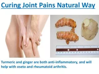
A 35-year-old lady presented with joint pains and low-grade fever for 6 months. She was previously diagnosed with rheumatoid arthritis and now has additional symptoms of decreased urinary output and vomiting. The clinical diagnosis is connective tissue disorder, possibly SLE with lupus nephritis or RA with nephritic/nephrotic syndrome. Autoimmune workup shows positive ANA, anti-dsDNA, and anti-Sm antibodies.

E N D
A YOUNG LADY WITH FEVER AND JOINT PAINS Dr. MUHAMMAD OSAMA REGISTRAR NEPHROLOGY, HFH,,Rwp.
Patient xyz 35 years of age R/O of Attock house wife by profession educated up-to middle married with 5 kids presented to us on 20th Dec with C/O • Join pains 6 months • Fever 6 months
PREMORBIDS NO PREMORBIDS
HOPI • Alright 6 months back, developed join pains started from • Small joints of both hand and feet • Red, swollen, painful, morning stiffness of 2-3 hours, restricted movements. • Movements improved with physical activity • After one month pain involved both wrist and elbow joints
Joint pain was associated with low grade fever that was intermittent with no spikes aggravated by physical activity relieved by antipyretics with no weight loss night sweats or anorexia.
After one month patient presented to PIMS • Diagnosed as a case of Rheumatoid arthritis(record N/A) • Treated with deltacortril, NSAIDS and methotrexate • After two months developed redness and itching on sun exposed skin in sun light, painless oral ulcers and red colored flat rash on cheeks and nose. • After 5 months of R.A treatment she presented to us with fever, joint pain, decreased urinary output and vomiting.
Urinary output decreased in the last one week in a progressive pattern with once daily voiding at the time of presentation • No change in color of urine • No burning micturition or flank pain
SYSTEMIC ENQUIRY • CVS : NOT SIGNIFICANT • RESPIRATION : NOT SIGNIFICANT • G.I : NOT SIGNIFICANT • CNS : altered sleep pattern and low mood 3 months • AUTOIMMUNE : BLUISH DISCOLORATION(INTERMITENT) OF FINGER TIPS 2 months • ENDOCRINE : NOT SIGNIFICANT • OBSTETRICAL : NO FETA LOSES, NO MISCARRIGES, NORMAL 30/7 CYCLE
PAST MEDICAL AND SURGICAL Hx • Not significant
FAMILY HISTORY • NO Hx OF RENAL OR CONNECTIVE TISSUE DISORDER
PERSONAL AND SOCIAL HISTORY • Married with five children 2 son and 3 daughters elder son 17 years and younger daughter 5 years • House wife • Educated up-to middle • No Hx OF cigarette, huqa smoking or alcohol intake
DRUG Hx • NSAIDS, deltacortril and methotrexate for 5 months • No Hx of blood transfusions • No Hx of tooth extraction
ANIMAL CONTACT • NOT SIGNIFICANT
TRAVEL Hx • NOT SIGNIFICANT
GENERAL PHYSICAL EXAMINATION • A middle aged lady with Height of 5.1 feet and weight of 55 kg lying comfortably in bed , well oriented in time and space with cannula passed in right hand • Pulse 92/min • B.p 140/80 • R.R 16 • Temp 99 F • Pallor absent • Cyanosis absent
Jaundice absent • Clubbing absent • Koilonychia absent • Splinter hemorrhage absent • Leuconychia absent • Osler’s node absent • Heberden’s node absent
Bouchard’s node absent • Palmar sweating absent • Palmar erythema absent • Dupuytren’s contracture absent • Proptosis absent • Periorbital,pedal edema present, pitting nature • Skin rash present: red flat rash continuous over cheeks and nose • Parotid gland not enlarged
Thyroid not palpable • Neck veins not engorged • Lymph node not palpable
SYSTEMIC EXAMNATION • CVS :- pulse: 92 R BP 14/80 JVP` pedal edema + Normal shape precordium with no scar Apex beat in 5th ICS medial to M.C line wit no thrill or other sounds Both heart sounds are of normal intensity, no added sounds or murmur • RESPIRATON:- R.R: 16 c no deformity scar or prominent veins noted Trachea central , apex beat in 5th ICS medial to M.C line, no tenderness, equal chest movements and vocal fremitus Upper liver border at 5th ICS, percussion note is resonant and equal on both sides B/L vesicular sound of normal intensity, no added sounds , B/L equal vocal resonance
G.I:- Painless oral ulcers on buccal mucosa, Flat abdomen with no visible peristalsis, central umbilicus, no scar striae or visible veins No rigidty of tenderness, no viscera or mass palpable No dullness or rigidty B.S 4/minute, no bruits friction or audible sounds SOFT NON TENDER BOWEL SOUNDS POSITIVE
CNS :- Fully conscious well oriented in time and space Speech : N C.N : N Motor system : N Sensory system : N Cerebellar signs : negative Signs of meningeal irritation : N Postural drop: negative
CLINICAL Dx • Connective tissue disorder most likely SLE with lupus nephritis • R.A with nephritic/nephrotic syndrome • cryoglobulinemia
LABS • BLOOD CP:- Tlc : 12.7k Hb : 10.1 with MCV of 88.4 and MCH of 29.2 Platelets : 178k ESR : 85 CRP : positive
LFTs : N • RFTs : - U: 234 Cr: 5.2
Serum electrolytes:- Na : 143 K : 5.6 Cl : 99 S.Ca: 7.2 (corrected 9.0 with S.albumin of 1.8)
URINE R/E :- Color : yellow Turbid PH 6.0 Albumin 2+ Sugar negative Sp gravity 1.025 Wbc 4-6 Rbc 15-20 Epithelial cast 1+
ABGs:- PH : 7.29 PCO2 : 30.2 HCO3 : 15.6
AUTOIMMUNE WORKUP • ANA : positive • Anti ds DNA : weak positive • Anti Sm : positive • C3 : 1.3 N (0.7-01.7) • C4 : 0.15 decreased (0.2-0.5) • RA factor : negative • Anti CCP antibodies : negative • Anti phospholipid antibodies : negative • Hepatitis B and C serology by ELISA : negative
HOSPITAL COURSE • With these findings we were suspecting SLE with RPGN/AKI • so we put the patient on hemodialysis and started with pulse dose of steroid followed by cyclophosphamide. • After one and a half week as patient became stable we did renal biopsy and the results were
After eleven sessions of hemodialysis and pulse steroid followed by oral steroids and Single dose of cyclophosphamide patient RFTs returned to normal in 8 weeks (U: 40 Cr: 1.2) • On the basis of clinical and lab findings our diagnosis is SLE with AKI/AIN or AIN sec to drugs.
LUPUS NEPHRITIS Literature review
DEFINITON • It is an immune complex GN that is a common and serious manifestation of SLE which itself is defined by a combination of clinical and lab features
EPIDEMIOLOGY Incidence and prevalence of SLE and LN is influenced by • Age • Gender • Ethnicity • Geographical area • Diagnostic criteria used
Peak incidence of SLE is 15-45 years of age • Women to men ratio is 10:1 • But LN effects both genders equally • LN is more severe in child and men • Additional risk factor for LN are younger age ,low social class, more ACR criteria for SLE, longer duration of disease, family history of SLE, Hypertension
ETIOLOGY • Etiology is influenced both by genetic and environmental factors • Multiple genes are involved, more than 17 loci have been identified for SLE development. • Concordance is observed in > 25% of identical twins and > 5% of fraternal twins • Def of C3 C4 and C1q is a important risk factor for SLE
PATHOGENESIS • Many autoantibodies are involved • In early stages there is defective clearance of apoptotic cells resulting in autoantigens production stimulating INF alpha and various inflammatory cytokines. • This result in production of auto antibodies • In LN auto antibodies found mostly are against ds DNA, Sm antigen, C1q, nucleosomes, • In general, Mesangial and sub-endothelial immune deposits are derived from deposition of circulating immune complexes.
For Sub epithelial immune complexes there is in situ formation • This immune complex deposition lead to complement activaiton and complement mediated damage, activation of procoagulant factors leading to glomerular damage.
CLINICAL MANIFESTATIONS • SLE can involve any organ • Disease course is characterized by flare and remission. • Outcome of the disease depends on whether the damage is of healing or non healing nature.
SCORING SYSTEMS • SLEDAI • BILAG • SLAM • SLICC-ACR
EXTRA RENAL MANIFESTAIONS • Non specific • Malaise • Low grade fever • Poor appetite • Weight loss • Patchy alopecia • Oral or nasal ulcers • Arthralgia and nonersovie arthritis • Raynaud’s phenomenon
Photosensitivity • Butterfly rash • Livedo reticularis upto 15% of the cases • Serositisupto 40% of the cases • Pulmonary hypertension sec to pulmonary emboli or I.V coagulation in association with APA • Miscarriages • Thrombocytopenias • Neuropsychiatric involvement