Download
1 / 24
260 likes | 521 Vues
บทที่ 10 การวิเคราะห์ธาตุส่วนผสมทางเคมี Chemical composition analysis. 1302 423 Industrial Materials Testing Assistant Professor Dr. Sukangkana Lee. วิธีการวิเคราะห์. XRF (X - ray fluorescence) XRD (X-ray diffraction) http://www.matter.org.uk/diffraction/x-ray/x_ray_diffraction.htm

E N D
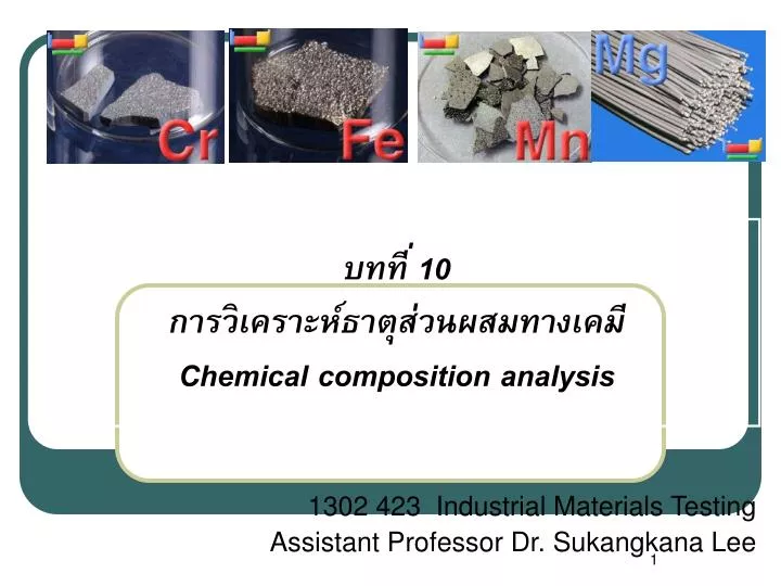
บทที่ 10 การวิเคราะห์ธาตุส่วนผสมทางเคมีChemical composition analysis 1302 423 Industrial Materials Testing Assistant Professor Dr. Sukangkana Lee
วิธีการวิเคราะห์ • XRF (X-ray fluorescence) • XRD (X-ray diffraction) http://www.matter.org.uk/diffraction/x-ray/x_ray_diffraction.htm • Energy dispersive Spectrometer ในวิชานี้จะอธิบาย XRF และ Emission Spectrometer
ทบทวน • โครงสร้างอะตอม มีลักษณะอย่างไร • ตารางธาตุ แสดงอะไร • X-rays คือ อะไร
โครงสร้างอะตอม e- N K L M
X-rays • X-radiation (composed of X-rays) is a form of electromagnetic radiation. • X-rays have a wavelength in the range of 10 to 0.01 nanometers, comparable to spacing between atoms/ions/molecules in crystal • corresponding to frequencies in the range 30 petahertz to 30 exahertz (30 × 1015 Hz to 30 × 1018 Hz) • energies in the range 120 eV to 120 keV. • They are shorter in wavelength than UV rays.
1. XRF (X-ray fluorescence) • XRF analysis เป็นเทคนิคที่สามารถวิเคราะห์ชนิดของธาตุ (Qualitative) และปริมาณธาตุ (Quantitative) • สามารถวิเคราะห์ได้ทั้งของแข็งและของเหลว • เป็นการทดสอบแบบไม่ทำลาย • สามารถวิเคราะห์ได้โดยไม่ต้องมีการเตรียมผิวมาก่อนก็ได้ • สามารถวิเคราะห์ธาตุที่มีความเข้นข้นน้อยๆประมาณ 5-500 ppm ได้
ชิ้นงานต้องมีเส้นผ่าศูนย์กลางประมาณ 10-50 mm และสูงไม่เกิน 50 mm ผิวหน้าชิ้นงานของแข็งต้องเรียบ และผ่านการขัดเงา • ถ้าเป็นชนิดผงต้องหนักอย่างน้อย 1 gram • ใช้เวลาวิเคราะห์แต่ละครั้งประมาณ 15 นาที
ข้อจำกัดคือ ไม่สามารถวิเคราะห์ธาตุที่มี atomic number น้อยกว่า 11 ได้ (H, He, Li, Be, B, C, N, O, F, Ne and Na) ตัวอย่างการวิเคราะห์ XRF • การวิเคราะห์เปอร์เซนต์ของธาตุผสมในโลหะผสม เซรามิกส์ แก้ว เป็นต้น • การวิเคราะห์ชนิดของธาตุซึ่งไม่สามารถวิเคราะห์ได้โดยวิธีอื่น
หลักการวิเคราะห์ • The process in which an x-ray is absorbed by the atom by transferring all of its energy to an innermost electron is called the "photoelectric effect."
Primary x-ray beam Ejected core electron M L K Electron from outer shell fills the hole Secondary x-ray beam หลักการวิเคราะห์ • เมื่อวัสดุได้รับการกระตุ้นจาก X-rays ที่มีพลังงานสูงมากพอที่จะกระตุ้นให้ electron (ชั้น K) หลุดออกมาจากวงโคจร(Ejected core e-) ก็จะทำให้อิเลคตรอนจากชั้นวงนอกเลื่อนลงมาเติมช่องว่าง โดยอิเลคตรอนชั้นนอกจะมีการปล่อยพลังงานส่วนหนึ่งออกมาในรูปของ secondaryX-ray
จำนวนพลังงานที่สามารถกระตุ้น core electron ให้หลุดออกมา (Primary x-ray) และ จำนวนพลังงานที่ปล่อยออกมา (Emitted secondary x-ray) จะเป็นค่าคงที่ของแต่ละอะตอม • XRF Titanium
L Mshell K Lshell Kshell K N • ถ้าอิเลคตรอนจากชั้น L shell ลงไปอยู่ชั้น K shell จะปล่อยพลังงานที่มีความยาวคลื่นค่าหนึ่งเรียกว่าKα • ถ้าอิเลคตรอนจากชั้น M shell ลงไปอยู่ชั้น K shell จะปล่อยพลังงานที่มีความยาวคลื่นค่าหนึ่งเรียกว่าKβ
X-ray K-series spectral line wavelengths (nm) for some common target materials.[13] ^David R. Lide, ed. CRC Handbook of Chemistry and Physics 75th edition. CRC Press. pp. 10–227. ISBN 0-8493-0475-X.
Specimen 1 2 Secondary x-ray beam X-ray filter Intensity 3 Primary x-ray beam X-ray detector X-ray source Energy (wavelength) • ยิง x-ray ที่มีพลังงานเพียงพอไปกระตุ้นชิ้นงานโดยตรง • Secondary x-ray ถูกปล่อยออกมาจากชิ้นงานไปสู่ x-ray detector • X-ray detector จะวัด ค่า energy wavelength ของ KและKเปรียบเทียบกับค่ามาตรฐาน
Intensity Energy (wavelength) Target X-ray detector 3 1 2 X-ray filter Secondary x-ray beam Primary x-ray beam Specimen X-ray source • ยิง x-ray ที่มีพลังงานเพียงพอไปกระตุ้น target ระยะซึมลึกประมาณ 10-4 ถึง 10-5m • Secondary x-ray จาก Target ถูกปล่อยไปสู่ Specimen และ สะท้อนไปสู่ X-ray detector • X-ray detector จะวัด ค่า energy wavelength ของ KและKเปรียบเทียบกับค่ามาตรฐาน
2. Optical Emission Spectrometer • เป็นการหาชนิด และปริมาณของธาตุ โดยใช้หลักการการกำเนิด X-ray โดยที่จะเป็นการวิเคราะห์พลังงานของ X-ray ที่ได้จากผิวชิ้นงาน • สามารถวิเคราะห์ 30 ธาตุได้ในเวลา 1-2 นาที
เมื่ออิเลคโตรดได้รับพลังงานไฟฟ้าจะให้กำเนิดลำอิเลคตรอน ด้วยกำลังประมาณ 800-100 V และตกกระทบผิวชิ้นงาน ผ่านบรรยากาศของแกสอาร์กอน • อิเลคตรอนในอะตอมของชิ้นงานที่มีระดับพลังงานต่ำสุด (เรียกว่า ground state )จะถูกกระตุ้นให้มีระดับพลังงานสูงขึ้น (เรียกว่า Excited state) อะตอมที่อยู่ในสภาวะนี้จะไม่เสถียร จึงพยายามลดพลังงานลงมา • จึงปลดปล่อยพลังงานส่วนเกินออกมาในรูปของแสง (Light Emission) หรือ คลื่นแม่เหล็กไฟฟ้า ที่มีความยาวคลื่นเฉพาะตัว
Emission of atom 2.Excitation 1.Ground state 3.Emission Supplied Energy +E e- คายพลังงานออกมาในรูปของแสงมีความยาวคลื่น e- Initial Energy, E1 Energy, E2 Emitting a Photon (E2-E1) การเปลี่ยนแปลงพลังงานของอะตอม (Atomic Phenomenon)
Planck’s equation • E คือ พลังงานที่แตกต่างกันระหว่าง 2 ระดับพลังงาน • h คือ ค่าคงที่ของ planck • คือ ความถี่ของรังสี • C คือ ความเร็วแสง • คือ ความยาวคลื่น ดังนั้น ค่าพลังงานจะแปรผกผันกับค่า ความยาวคลื่น แต่ละธาตุจะมีชุดความยาว คลื่นที่เกิดจากการเปล่งพลังงาน (Emission) เฉพาะตัว
แสงที่เกิดขึ้นจะถูกส่งผ่านไปยังระบบแยกความยาวคลื่นแสง ซึ่งเรียกว่า Spectrometer ซึ่งมีลักษณะเป็นเลนส์นูน และปริซึมสามเหลี่ยม (ผลึกของ silicon or Lithium)เพื่อให้เกิดการหักเหของแสง เป็น Spectrum • จากนั้นแสงที่ถูกแยกความยาวคลื่นแล้วจะถูกส่งไปยัง Detectorเพื่อเปลี่ยนความเข้มแสงให้เป็นสัญญาณไฟฟ้า และระบบวิเคราะห์สัญญาณไฟฟ้าให้เป็นความเข้มข้นของธาตุ • โดยเปรียบเทียบกับสัญญาณของมาตรฐานที่เราทราบความเข้มข้นที่ได้ทำการวิเคราะห์ และบันทึกไว้ก่อนหน้านี้
แผนผังการทำงานของ Spectrometer Detector Computer software Work Table Sample Discharge Spectrometer Light Emission Chamber (with argon flow) Electrode
Work Table Spectrometer