Download
1 / 3
30 likes | 131 Vues
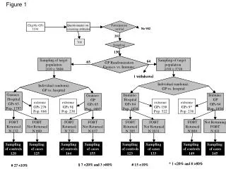
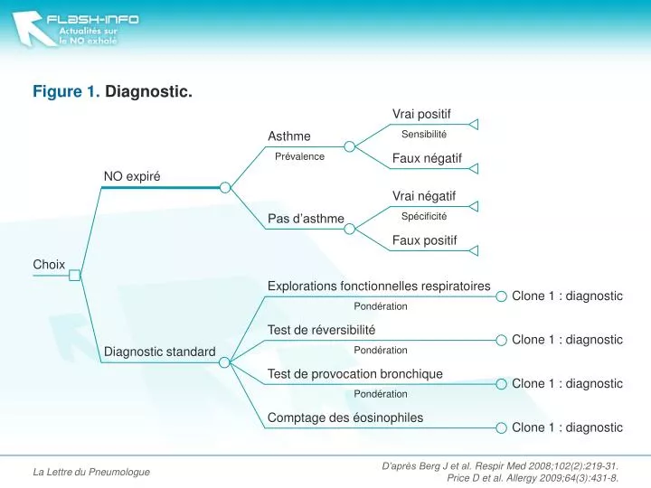
Figure 1. Diagnostic. Vrai négatif. Faux négatif. Vrai positif. Faux positif. Sensibilité. Spécificité. NO expiré. Diagnostic standard. Pas d’asthme. Asthme. Prévalence. Choix. Comptage des éosinophiles. Explorations fonctionnelles respiratoires. Test de réversibilité.

E N D
Figure 1. Diagnostic. Vrai négatif Faux négatif Vrai positif Faux positif Sensibilité Spécificité NO expiré Diagnostic standard Pas d’asthme Asthme Prévalence Choix Comptage des éosinophiles Explorations fonctionnelles respiratoires Test de réversibilité Test de provocation bronchique Clone 1 : diagnostic Clone 1 : diagnostic Clone 1 : diagnostic Clone 1 : diagnostic Pondération Pondération Pondération La Lettre du Pneumologue D’après Berg J et al. Respir Med 2008;102(2):219-31. Price D et al. Allergy 2009;64(3):431-8.
Figure 2. Suivi. Asthme bien contrôlé Asthme modéré Hospitalisation NO expiré Recommandations standard Exacerbation Asthme sévère Pas d’hospitalisation Choix La Lettre du Pneumologue D’après Berg J et al. Respir Med 2008;102(2):219-31. Price D et al. Allergy 2009;64(3):431-8.
Figure 3. Résultats. La Lettre du Pneumologue D’après Berg J et al. Respir Med 2008;102(2):219-31. Price D et al. Allergy 2009;64(3):431-8.