Download
1 / 32
420 likes | 708 Vues
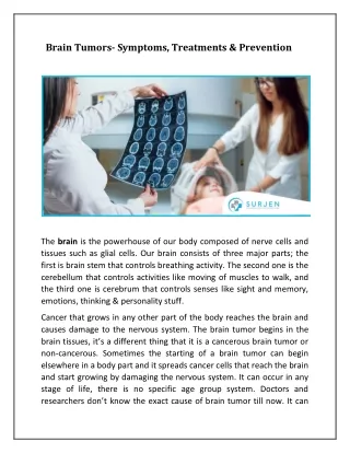
BRAIN TUMORS. Elshami Elamin, MD. Introduction. Intracranial neoplasms arise from: Brain cells Meninges Pituitary gland Skull Residual embyronic tissue Mets from lung, breast etc… Most common solid tumors in children 2 nd cause of cancer death in chlidren. Symptoms, Signs. Headache

E N D
BRAIN TUMORS Elshami Elamin, MD
Introduction • Intracranial neoplasms arise from: • Brain cells • Meninges • Pituitary gland • Skull • Residual embyronic tissue • Mets from lung, breast etc… • Most common solid tumors in children • 2nd cause of cancer death in chlidren
Symptoms, Signs • Headache • N/V • Papilledema • Lateralizing signs; • Hemiparesis • Aphasia • Visual-field deficits • Seizures • Altered Mental Status
Diagnosis • MRI; (gold standard) • To define number of met nodules • Leptomeningeal dz • High G or malignant tumors are enhancing masses in the white matter surrounded by edema • Low G gliomas are nonenhancing • Magnetic Resonance Spectroscopy: • Differentiate between low and high G gliomas • CT scan • PET scan (75% sensitivity, 83% specificity)
PATHOLOGY • Gliomas arise from: • Astrocytes • Oligodendrocytes • Ependymomas: • Low G histologically • High recurrence rate • Medulloblastomas: • High G • Primitive Neuroectodermal Tumors (PNETs): • High G • Pineoblastomas, neuroblastomas • Extra-axial • Meningiomas
GRADING of GLIOMAS • Based on endothelial proliferation, cellular pleomorphism, mitosis, necrosis. • High Grade: • GBM • Anaplastic astrocytoma • Anaplastic oligodendroglioma • Gliosarcoma • Low Grade: • Astrocytoma • Oligodendrogioma • Mixed oligoastrocytoma
STAGING • Not applicable in primary Brain tumors: • Locally invasive • Do not spread to LN or distant organs • Enhanced complete spinal MRI and CSF eval: • Medulloblastoma • Ependymoma • PENT
PROGNOSIS • With conventional treatment: • Anaplastic astrocytoma: MS is 3 Yrs • GBM: MS is 1 Yr • Low G gliomas: MS is 5-10 Yrs • Pts >40Yrs: MS is < 5 Yrs • Brain mets: MS 4-6 m • Single met treated with surgery/G-K: MS > 40Wks
TREATMENT • Supportive: • Anticonvulsant • Seizure at presentation • Prophylaxis only during peri-operative period • Steroids; • Most pts stop steroids when complete RT • If steroids >6 wks: • PCP prophylaxis • Definitive:
Surgery • Goals of surgery: • histologic diagnosis • reducing tumor burden and associated mass effect • maintaining or reestablishing pathways for CSF flow • achieving potential “cure” by gross total removal
RT • Brachytherapy • Radiosurgery • Gamma Knife • iodine-131–labeled antitenascin monoclonal antibody
Adult Low-Grade Infiltrative SupratentorialAstrocytoma/Oligodendroglioma
Management • MRI compatible with primary brain tumor: • Maximal safe resection • Feasible: • post-op MRI • >45Y; Observe or Post-op RT • <45Y; observe • Not feasible: • Stereotactic or open biopsy • Post-op observe, RT, or Chem • F/U: MRI q3-6m x 5Yrs • Recurrence: • Surgery, RT, Chemo
low-grade gliomas • complete surgical resection of hemispheric astrocytomas • Incomplete resection: • Astrocytoma: Post-op RT • oligodendrogliomas or mixed gliomas: may benefit from chemotherapy • Recurrent unresectable dz • RT
Adult Intracranial Ependymoma or Anaplastic Ependymoma • MRI/CT compatible with primary brain tumor: • Maximal resection: (Stereotactic or open biopsy if not feasible) • Postop staging and Adj therapy: • Negative post-op MRI brain,spine +/- CSF • Limited-field RT • Positive post-op MRI brain, spine +/- CSF • Craniospinal RT • F/U: • MRI/CT q3-6mx2Y, then q6-12m • Recurrence: • Resection, RT or Chemotherapy or supportive care
Anaplastic Astrocytoma/AnaplasticOligodendroglioma/ Glioblastoma Multiforme
MRI suggestive of high-G glioma: • Multidisciplinary planning • Maximal Resection • Feasible • Max excision +/- BCNU wafer • Not feasible (Stereotactic or open biopsy)
Adjuvant Therapy • Anaplastic astrocytoma or Anaplastic oligodendroglioma: • Adj RT +/- Chemo • Glioblastoma Multiforme (GBM): • s/p BCNU wafer: • Adj RT ± chemotherapy • No BCNU wafer: • Adj RT ± concurrent and adj temozolomide
High G Gliomas • Post-op RT: • Involved-field 60Gy prolong Survival • 50% of anaplastic astrocytomas respond • 25% of GBM respond • Complete response is rare
High G Gliomas • Adj chemo + RT prolong survival: • BiCNU • PCV • Temozolomide • GBM: Adj Temodar + RT standard of care
Chemotherapeutic regimens for gliomas • Single-agent BiCNU • BiCNU 200 mg/m2 IV q8wk • (maximum cumulative dose,1,500 mg/m2) • Single-agent temozolomide • Temozolomide 150-200 mg/m2 PO on days 1-5 • Repeat cycle every 28 days. • Standard PCV • Procarbazine 60 mg/m2/d PO on days 8-21 • Lomustine (CeeNu) 110 mg/m2 PO on day 1 • Vincristine 1.4 mg/m2 IV on days 8 and 29 • (maximum dose, 2 mg) • Repeat cycle every 6-8 weeks, optimally for 6 cycles. • Intensified PCVa • Procarbazine 75 mg/m2/d PO on days 8-21 • Lomustine (CeeNu) 130 mg/m2 PO on day 1 • Vincristine 1.4 mg/m2 IV on days 8 and 29 • (no dose limit) • Repeat cycle every 6 weeks.
High G Gliomas F/U • MRI 2-6 wk after RT • Then every 2-3 m x 2-3 y
High G Gliomas F/U • MRI 2-6 wk after RT: • Then every 2-3 m x 2-3 y • Recurrence: • Diffuse, Multiple: • Best supportive care if poor PS • chemo • Surgery for symptomatic, large lesions • Local: • Resectable: surgery +/- BCNU polymer, RT, chemo • Unresectable: RT, chemo
Limited (1-3) mets • SCLC or Disseminated systemic dz: • WBRT • Limited systemic disease: • Surgery or Stereotactic radiosurgery • consider WBRT • If unresectable; WBRT and/or radiosurgery
>3 Metastatic Lesions • WBRT ± stereotactic radiosurgeryc
Leptomeningeal disease • DIAGNOSIS: • Positive CSF cytology OR • Positive MRI with supportive clinical OR • CSF findings in a pt known to have cancer OR • Signs and symptomswith supportive CSF in apt known to have amalignancy
Leptomeningeal diseaseTreatment • Poor risk: • Palliative RT, supportive care • Good risk: • IT chemo • Palliative RT to symptomatic/bulky sites • Systemic chemo
Leptomeningeal disease • CSF flow scan: • Normal flow: • Induction IT chemo x 4-6wks • Recheck CSF • Abnormal flow: • RT to site of obstruction +/- IT chemo • Repeat CSF flow scan
Leptomeningeal diseasePost-induction Therapy • Negative CSF: • Maintenance IT chemo • Monthly CSF • Positive CSF: • Clinical leptomeningeal dz; • Supportive care, RT, chemo • No Clinical leptomeningeal dz; • Continue IT chemo • CSF continues to be +ve: • Supportive care
NonimmunosuppressedPrimary CNS Lymphoma • CT/MRI suggestive of lymphoma: • Hold steroids, if possible • CSF, Biopsy • Slit lamp eye exam • HIV test • Spinal MRI
NonimmunosuppressedPrimary CNS LymphomaTREATMENT • Good PS, CrCl > 50: • High-dose MTX-based +/- RT • LP or spinal MRI +ve, • consider IT chemo • Poor PS, CrCl < 50: • Whole-brain RT (45 Gy) or Chemotherapy • Eye exam +ve • RT to orbits • LP or spinal MRI +ve, • consider IT chemo + focal spinal RT
Metastatic Spine Tumors • Asymptomatic: • Surgery or RT or Chemo or Observation • Symptomatic (Pain, Neuro): • Steroids if abnormal neuro exam • Spinal MRI: • Spinal Cord Compression: • Surgery if: • Spinal instability • Radioresistant • Rapid neuro deterioration • Unknown primary • Previous RT • RT • Chemo • No Spinal Cord Compression: • RT, Vertebroplasty, Surgery if spinal instability