Download
1 / 2
20 likes | 488 Vues
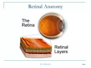
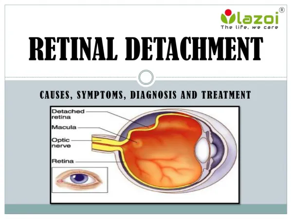
Retinal detachment is when the retina – a thin membrane of nerve tissue found in the eye – comes off the back of the eye, either partially or entirely, causing blurring or lost vision. Treatment for this kind of condition should be undertaken as soon as possible, as retinal detachment surgery is the only way to prevent serious complications.

E N D
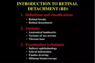
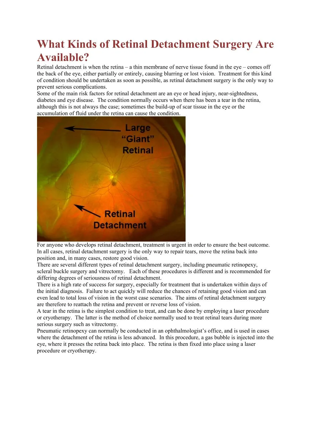
What Kinds of Retinal Detachment Surgery Are Available? Retinal detachment is when the retina – a thin membrane of nerve tissue found in the eye – comes off the back of the eye, either partially or entirely, causing blurring or lost vision. Treatment for this kind of condition should be undertaken as soon as possible, as retinal detachment surgery is the only way to prevent serious complications. Some of the main risk factors for retinal detachment are an eye or head injury, near-sightedness, diabetes and eye disease. The condition normally occurs when there has been a tear in the retina, although this is not always the case; sometimes the build-up of scar tissue in the eye or the accumulation of fluid under the retina can cause the condition. For anyone who develops retinal detachment, treatment is urgent in order to ensure the best outcome. In all cases, retinal detachment surgery is the only way to repair tears, move the retina back into position and, in many cases, restore good vision. There are several different types of retinal detachment surgery, including pneumatic retinopexy, scleral buckle surgery and vitrectomy. Each of these procedures is different and is recommended for differing degrees of seriousness of retinal detachment. There is a high rate of success for surgery, especially for treatment that is undertaken within days of the initial diagnosis. Failure to act quickly will reduce the chances of retaining good vision and can even lead to total loss of vision in the worst case scenarios. The aims of retinal detachment surgery are therefore to reattach the retina and prevent or reverse loss of vision. A tear in the retina is the simplest condition to treat, and can be done by employing a laser procedure or cryotherapy. The latter is the method of choice normally used to treat retinal tears during more serious surgery such as vitrectomy. Pneumatic retinopexy can normally be conducted in an ophthalmologist’s office, and is used in cases where the detachment of the retina is less advanced. In this procedure, a gas bubble is injected into the eye, where it presses the retina back into place. The retina is then fixed into place using a laser procedure or cryotherapy.
The next type of procedure is the scleral buckle, which is done by placing a flexible band around the eye with the aim of counteracting the force that is pulling on the retina and causing it to detach. Any fluid behind the retina will be drained away, and the retina should return to its normal place at the back of the eye. This type of retinal detachment surgery is more complicated, and therefore is normally carried out in a surgery clinic or a hospital operating room. Both local and general anaesthesia may be used. The most serious cases of retinal detachment are treated with a type of surgery known as vitrecto my, which usually takes place in a surgical clinic with he use of local anaesthetic. In this procedure, the vitreous gel is removed from the centre of the eye as it allows the surgeon better access to the retina. After this has been done, the surgeon may treat the tear in the retina, remove any scar tissue and flatten areas where the retina has become detached from the back of the eye. As with pneumatic retinopexy, a bubble of gas or oil is injected into the eye to push the retina into place until the detachment has healed. All of these procedures are necessary to treat retinal detachment, as surgery is the only way to fully treat this condition and prevent vision from progressively worsening. In all cases, a reputable ophthalmologist can best advice on the right treatment option for retinal detachment issues, based on the severity of the condition and any other eye issues.