Download
1 / 80
980 likes | 2.2k Vues
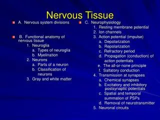
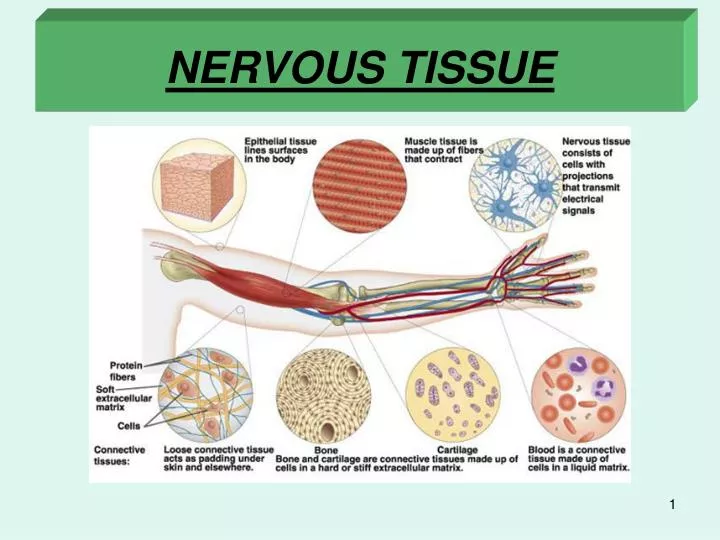
NERVOUS TISSUE. Units of morphology. Organ system. Ectoderm. Nervous tissue. Organ. Endoderm. Epithelial tissue. Tissue. Mesoderm. Connective tissue. Cell. Muscle tissue. Four basic tissue. proper C.T. Blood C.T. Cartilage. Bone. Adipose Tissue. Introduction.

E N D
Units of morphology Organ system Ectoderm Nervous tissue Organ Endoderm Epithelial tissue Tissue Mesoderm Connective tissue Cell Muscle tissue
Four basic tissue proper C.T. Blood C.T. Cartilage Bone Adipose Tissue
Introduction • There are two basic systems of internal communication and physiological homeostasis in the body: the endocrine system and the nervous system. • The nervous system is derived from embryonic neuroectoderm. • The human nervous system is divided anatomically into: 1-Central Nervous System (CNS), consisting of the brain and spinal cord. 2-Peripheral Nervous System (PNS), consisting of nerve fibers, aggregates of nerve cells and glia and ganglia. • It is estimated that the human nervous system consists of at least 10 billion neurons. • Nervous tissue consists of two groups of cell types: • 1-Nerve cells (Neurons) • 2-Neuroglia.
Central Nervous System (CNS) • The nerve cell bodies (perikarya) of the CNS are often found in groups ("nuclei"). -The brain and spinal cord are composed of gray matter and white matter. • Gray matter contains nerve cell bodies (perikarya), neuroglia, and a complicated network of cell processes (neuropil). • White matterlacks nerve cell bodies (perikarya), but has many processes of neurons. The white appearance is the result of the myelin that envelops many of the neuronal processes. Neuroglia are also found in the white matter and the nuclei seen in white matter belong to neuroglia. -In the Peripheral Nervous System (PNS), perikarya are found only in ganglia (apart from in some sensory regions such as the retina and olfactory mucosa).
BRAIN ANATOMY Cerebrum Cerebral Hemispheres (silver stain) gyri sulci gyrus sulcus Cerebellum Spinal Cord grey white matter matter
CENTRAL NERVOUS SYSTEM Brain Cortex (grey matter) Nuclei Medulla (cell bodies) (white matter) brain stem spinal cord golgi type I neuron white matter (axon tracts) golgi type I neuron golgi type II grey matter neurons (cell bodies)
SPINAL CORD dorsal root ganglia dorsal root muscle sensory stretch white receptor dorsal matter horn grey receptor matter meninges motor ventral end plates horn motor ventral root blood central canal vessel white matter grey matter
Neurons • Neurons are post-mitotic structures that shortly after birth lose the ability to divide. • Further changes involve only reduced number of neurons (neuronal death), or changes in volume or in neuronal connections. • Neurons have two special properties: 1-Irritability (the ability to respond to a stimulus) 2-Propagation of impulses (the ability to conduct impulses). • The morphofunctional unit of the nervous system is the neuron. • Similar to the Cell Theory, which stipulates the cell as the basic building block of the body, the Neuron Theory describes the neuron as the basic building block of the nervous system, and that the nervous system functions through transmission of information through networks of neurons.
Most neurons have three main parts: 1-Dendrites 2-Perikarya (cell bodies) 3-Axon • The dendrites are receptive to stimuli and bring stimuli from the environment (sensory epithelial cells or other neurons) to the cell body. There are usually several dendrites per neuron. • The perikaryon (cell body) is also receptive to stimuli, but also serves as the trophic or synthesizing center for the whole nerve. • The axon is a long process emerging from the cell body. There is only a single axon for each neuron. The axon transmits impulses to other neurons, or to effectors: muscle or gland cells. The distal portion of the axon is usually branched (terminal arborization). • Neurons and their processes are very variable in form and size. Some neurons are very large (with perikarya of up to 150m), whereas others are very small (perikarya of only 4-5m).
ANATOMY OF A NEURON impulse conduction axons dendrites axo-somatic synapse Nissl’s substance axo-dendritic synapse boutons axon axon hillock axo-axonic synapse myelin sheath oligodendrocyte unmyelinated collateral myelin sheath central nervous system (CNS) myelin sheath peripheral nervous systen (PNS) Schwann cell motor end plate synaptic vesicles motor end plate
MOTOR NEURON CELL BODY (H&E) DENDRITIC SPINES (boutons)-silver axo-denritic synapses dendrite (boutons) Nissl’s substance nucleolus dendrite nucleus axon
Morphological classification of neurons • Neurons are classified according to the size, number and shape of their processes. 1-Unipolar (pseudounipolar) neurons have a single process (axon). These are found in sensory ganglia of dorsal roots of spinal nerves. 2-Bipolar neurons have two processes (one dendrite and one axon). These are very rare and have a limited distribution in the body. They are present in special sensory structures including the retina, olfactory epithelium, and vestibular and cochlear nerves). 3-Multipolar neurons possess several processes (several dendrites and a single axon). Most neurons belong to this category.
The axons of small diameter are usually non-myelinated fibers, whereas the thicker axons have concentric wrappings of the enveloping cell to form the myelinated sheath. • The fibers with myelinated sheaths are called myelinated fibers. • Myelinated nerves, composed mainly of myelinated axons, appear white in the fresh state. • The sheath of myelinated fibers is formed by concentric layers of membranes of the Schwann cell (or oligodendrocyte in the CNS) around the axon, which unite to form a lipoprotein complex. • This stains black with osmium tetroxide. • The whorled structure of the myelin sheathe when examined by transmission electron microscopy is seen as a repeating dark line (major dense line) and a thinner repeating intraperiod line. • The major dense line is formed by the fusion of two of the inner layers of sheath cell membrane, whereas the intraperiod line is formed by the fusion of the outer layers of sheath cell membrane when they come in contact as a result of the concentric arrangement. • The myelin sheath is essentially an accumulation of closely packed whorls of lipoprotein rich membranes surrounding the axon.
Physiological classification of neurons • Neurons may also be classified according to their function. 1-Sensory neurons. These receive sensory stimuli from the environment (from receptors) and from within the body (e.g. unipolar neurons). 2-Motor neurons. These control the effector organs (muscles, exocrine glands, endocrine glands) 3-Interneurons (Intermediate neurons). These are typically found in the CNS and connect between other neurons (often between sensory and motor neurons). 4-Neurosecretory neurons. These are specialized neurons that synthesize and secrete hormones.
NEURON TYPES Bipolar Neuron (retina & olfactory epithelium) Pseudounipolar Neuron (sensory spinal reflex arc) Unipolar Neuron (embryonic)
PERIPHERAL NERVE ENDINGS-(MUSCLE) visceral visceral sensory (proprioreceptors) motor motor unit Neuromuscular Spindle motor end plates (proprioreceptors (boutons)
Each neuron has 3 physiological parts or segments: 1-Receptive segment (dendrites and perikaryon). The perikaryon also has an additional trophic and synthesizing role. 2-Conductive segment(axon) 3-Transmissive segment(synapse).
Reflex arcs • The functional roles of various neurons are best illustrated by simple reflex arcs in which peripheral receptors are connected to peripheral effectors in a neuronal network. 1-Stimulation of the receptor (in skin or skeletal muscle spindle) 2-Propagation of an impulse via afferent sensory nerve (unipolar neuron), which enters the gray matter of the spinal cord. 3-Interneurons connect with cell body of motor neuron in ventral horn. 4-Motor neuron transmits the efferent impulse to an effector. 5-The effector e.g. motor end plate of skeletal muscle responds to the impulse. • (An analogy can be made to a cable system of telephone wires. The phone message is sent to a central exchange, that directs the message to the correct connection, resulting in a specific response by the recipient).
MORPHOLOGY OF NEURONS (1)DENDRITES • Most nerve cells have several dendrites. • These increase the receptive area of the neuron. • Dendrites do not maintain a constant diameter (unlike axons) and transmit impulses to the cell body decrementally (unlike axons). • The regions of the dendrites closest to the perikaryon are usually larger, than those farther away. • Typically dendrites have large numbers of thorny spines, which are now known to be areas of synaptic contact.
2- PERIKARYON The perikaryon (neuronal cell body or soma) consists of the nucleus and surrounding cytoplasm. (The term perikaryon implies the area surrounding the nucleus, but the term is used freely today to describe the whole cell body including the nucleus). The perikaryon is the trophic center of the neuron involved in protein synthesis. The surface of the perikaryon receives nerve impulses and is the site of many synapses, bringing excitatory or inhibitory stimuli.
Nucleus • The nuclei of perikarya are large, regular, round or oval, typically situated fairly centrally. The nuclei are euchromatic (pale staining with dispersed chromatin). Such large regular nuclei are typical of cells involved in intense synthetic activities. Sex chromatin (Barr’s body) is commonly seen in the nuclei of females. Rough Endoplasmic Reticulum (RER) - Nissl bodies • RER is abundant in the cytoplasm and is associated with the protein synthetic activities of the neurons. The RER is basophilic as seen in regular (H&E) staining by light microscopy. This RER is also stained with cresyl violet in the Nissl staining technique (Nissl bodies). In the event of injury to axons, Nissl bodies, are displaced to the periphery of the perikaryon. Golgi bodies • Large well developed Golgi bodies are present in the perikarya. Mitochondria • Many large mitochondria are found throughout the perikaryon. Neurofibrils • Neurofibrils are seen in perikarya (and also in the nerve processes) after silver impregnation techniques. At the electron microscope level these are seen to consist of clumped neurofilaments and neurotubules. Lipofuscin • Lipofuscin is a brown pigment that is common in perikarya of aged neurons. It is now known to be common to post-mitotic cells and to consist of large secondary lysosomes.
NERVE CELL BODY myelinated axon axo-denritic synapse unmyelinated axons axo-somatic synapse axon hillock neurotubules neurofilaments axon vesicles (neurotransmitters) mesaxon axon
AXONS • Each neuron has a single axon. • The diameter of the axon is fairly constant. • The length of axons is fairly variable, and some reach up to 100 cm (the axons innervating the toes have their cell bodies in the spinal cord). • All axons originate in a short pyramid-like structure called the axon hillock, which lacks Nissl substance. • The plasma membrane of the axon is termed the axolemma, and the cytoplasm of the axon is termed the axoplasm. • In myelinated axons the initial portion, between the axon hillock and the start of the myelin sheath, is called the initial segment. • Axons sometimes have right-angled branches known as axon collaterals. • The nerve impulse travels down the axon non-decrementally.
Myelinated fibers • Nerve fibers consist of axons enveloped by special sheaths. • In peripheral nerves the sheath cell is the Schwann cell, whereas in the CNS, the sheath-forming cells are the oligodendrocytes.
MYELIN MESAXON MYELIN SHEATH Schwann cell AXON cytoplasm major dense interperiod line
MYELINATED AXONS Myelin Sheath Schwann Cell node of Ranvier axon
SHEATH OF SCHWANN node of Ranvier internodal region clefts of collateral Schmidt-Lanterman Schwann sheath axon
If a single fiber of a myelinated peripheral nerve is teased, stained with osmium tetroxide and examined by light microscopy, the myelin sheath surrounding the axon is seen as a series of myelinated internodes (0.08-1.00 mm) separated by nodes of Ranvier. • (The myelinated axon is some what similar to a long string of sausages). • The myelin of each internode is formed by a single Schwann cell, whose nucleus is seen at the periphery. • Tangential non-stained areas (similar to arrow heads) are seen in the myelin of the internodes (Schmidt-Lantermann clefts). • These are areas of cytoplasm of the Schwann cells, where the membranes are not closely apposed. • An endoneurial connective tissue sheath surrounds each fiber.
In wax sections stained with H & E, the lipid of the myelin is dissolved by the xylene or chloroform during processing and the site of the myelin sheath appears empty apart from a fine network stained by the eosin. This is known as neurokeratin. • Myelinated axons of the CNS have myelin sheaths, similar to those of the peripheral nerves. However, a single oligodendrocyte produces the myelin sheaths of several axons. No endoneurial connective tissue sheath is present. The nodes of Ranvier are larger and exposed to the extracellular space.
Nodes of Ranvier • The nodes of Ranvier have several important features: a) sites of axon collaterals b) large concentrations of mitochondria in the axon at these sites (high local metabolic activity) C) site of saltatory conduction (non- decremental) D) sites of paranodal loops (important in the saltatory conduction).
NODE OF RANVIER AND CLEFTS OF SCHMIT-LANTERMAN Myelin Axon Cleft of Sheath Schmidt-Lanterman Node
Axonal transport • Transport of molecules along the axon (axonal transport) is in two directions: 1- anterograde (from the cell body to the terminal synapse) or 2- retrograde (in the direction of the cell body). The axonal transport involves neurotubules and neurofilaments. Two different systems of axonal transport occur: 1- Slow axonal transport system, from the cell body in a single direction at a rate of about 1mm per day. This system conveys components needed for growth and regeneration of the axon. 2- Fast axonal transport system, which occurs in both directions, at a rate of about 100-200 mm per day. This system involves transport of enzymes needed for synthesis of neurotransmitters within the terminal synapse.
NEUROGLIA Glia or neuroglia get their name from the Greek word for "glue". There is very little connective tissue in the CNS, and the structural support for neurons comes from neuroglia and their processes. • It is estimated that for every neuron there are at least 10 neuroglia, however, as the neuroglia are much smaller than the neurons they only occupy about 50% of the total volume of nerve tissue. • Neurons cannot exist or develop without neuroglia. There are 4 basic types of neuroglia, based on morphological and functional features. 1- Astrocytes (or Astroglia) 2- Oligodendrocytes (or Oligodendroglia) 3- Microglia 4- Ependymal cells • The astrocytes and oligodendroglia are large cells and are collectively known as Macroglia.
NEUROGLIA brain ventricles satellite central cell canal of spinal cord pseudounipolar ependymal neuron cerebro- spinal Cells fluid satellite cells Pseudounipolar neuron of peripheral ganglia
NEUROGLIA (nerve glue) Macroglia Protoplasmic Astrocyte (grey matter) Foot blood vessel Process capillary (pedicle) Fibrous Astrocyte (white matter) Foot Process blood vessel (pedicle) capillary
NEUROGLIA MICROGLIA (mononuclear phagocytic system) OLIGODENDROGLIA (forms Scwann sheath of CNS)
GREY AND WHITE MATTER Grey Matter WHITE AND GREY MATTER nerve cell bodies nerve white matter glial fibers nuclei White Matter dura axons grey matter mater (fibers) arachnoid membrane motor glial neurons pia nuclei mater
ASTROCYTES AND FOOT PROCESSES BLOOD VESSEL ASTROCYTE FOOT PROCESS
Neuroglia differ from neurons: 1 - Neuroglia have no action potentials and cannot transmit nerve impulses 2 -Neuroglia are able to divide (and are the source of tumors of the nervous system) 3 -Neuroglia do not form synapses 4 -Neuroglia form the myelin sheathes of axons.
Astrocytes (Astroglia) • These are present only in the CNSandare the largest of the neuroglia. They have many long processes, which often terminate in "pedicels" on blood capillaries and contribute to the blood-brain-barrier. There are two categories of astrocytes: A )Protoplasmic astrocytes. These are present in the gray matter of the brain and spinal cord. Their processes are relatively thick. B )Fibrous astrocytes. These are present in the white matter of the CNS. Their processes are much thinner than those of the protoplasmic astrocytes. • Because of their number and their long processes, the astrocytes appear to be the most important supporting elements in the CNS.
Oligodendrocytes • These are smaller than the astrocytes, with fewer and shorter processes. • They are found in both the gray and white matter of the CNS and are responsible for the formation of the myelin sheath surrounding axons. • The Schwann cells of the PNS belong to the oligodendrocytes and form the myelin sheath around peripheral axons.