Download
1 / 14
140 likes | 333 Vues
Red Cells. Prof. K. Sivapalan. ERYTHROCYTE- RBC. Biconcave disc. 7.2 μ x 2.2 μ No nucleus. PCV – 45, 35 % Hb% - 14.5 g/dL. - Males, 13.5 g/dL. - females Red cell count 5,000,000 / mm 3 . (5 x 10 6 ). Hemoglobin. 4 Units- Heme and peptide. 2 x α chains- 141 AA.

E N D
Red Cells Prof. K. Sivapalan
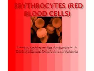
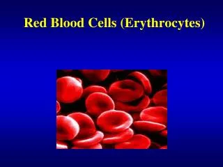
ERYTHROCYTE- RBC • Biconcave disc. • 7.2 μ x 2.2 μ • No nucleus. • PCV – 45, 35 % • Hb% - • 14.5 g/dL. - Males, • 13.5 g/dL. - females • Red cell count • 5,000,000 / mm3. (5 x 10⁶) Red Cells
Hemoglobin. • 4 Units- Heme and peptide. • 2 x α chains- 141 AA. • 2 x β chains- 146 AA. • Molecular weight- 67,000. • Hb A- α2β2. • Hb A2- α2δ2. (10 AA differ) • Hb F - α2γ2. (37 AA differ) • Hb S. (sickle cells) Red Cells
Heme. • Heme is attached to N terminals in the Peptide Chain. • Oxygenation is loose attachment to iron atom. Red Cells
Reaction of Heamoglobin. • 1 gram of hemoglobin binds to 1.34 ml of oxygen. • Hb4 + 4 O2 = Hb4O8 (Oxygenation) • Oxyhemoglobin – red color. • Deoxyhemaglobin- blue color - Cyanosis. • Carboxy hemoglobin- cherry red. • Methemoglobin- brown. Red Cells
Abnormalities of hemoglobin. • Hemoglobinopathies: Mutant genes → abnormal polypeptide chains. • Thalassemias: α or β thalassaemia - respective chain is absent or decreased. Defects in regulatory portion of the genes. Red Cells
HEMOPOISIS. • Stem cell. • Proerythroblast. • Basophelic. • Early normoblast. • Intermediate normoblast. • Hemoglobin. • Late normoblast. • Reticulocyte. • Erythrocyte. Red Cells
Hemopoisis. • Embryo- yalk sack. • Later- liver, spleen and lymph nodes. • Adults-redbone marrow. • Regulation: • Ischemia to kidney tissue → Erythropoitin by Juxta Glomerular Apparatus → increase in hemopoisis.Duration: 4 days. • Testesteron stimulates red cell production and result in increased Haemoglobin concentration in adult males. • Glucocorticoids also stimulate- polycythaemia in excess secretion. Red Cells
Limitations to Hemopoisis. • Protein- severe malnutrition. • Iron (Fe): Inclusion in haemoglobin. • Difficulty in absorption. • Food habits. • Loss [females]. • Cyanocobalamine (Vit. B12) and, Folic acid: • DNA synthesis. Red Cells
Fate of red cells. • Average life span – 120 days [ of the red cells, 1/120 destroyed daily]. • Destroyed by the Macropharges in spleen. • [Reticulo - Endothelial System] • Trabeculae in spleen – 3 μ. • Protein → Amino acid pool. • Heam →Iron + Porphyrin. • Iron → storage + recycled. • Porphyrin → Bilirubin – excreted. Red Cells
Bilirubin • Bound to albumin, taken up by the liver. [yellow] • Biliverdin – green. • Conjugated with glucuronic acid → water soluble and Excreated in bile – green. • Intestinal bacteria – stercobilinogen →absorbed – urobilinogen. Red Cells
Investigations on red cell. • Osmotic Fragility. • Two drops of blood in NaCl solutions with varying osmolality and observe for hemolysis. Spherocytosis- fragility increased. • Hemoglobin concentration. • Talqivist method. • Sahli method (acid haematin formation). • Calorimeters. • Oxygen content. • Pack cell volume. • Centrifugation. (buffy coat) • Red cell count. • Red cell pipette and hemocytometer. Red Cells
Red cell parameters. • Mean Corpuscular Hemoglobin [MCH] = Hb in g in 100 ml / No. of RBC in 100 ml. = 15/5x106 x 105 = 15/5x1011 = 30 x 10-12. = 27 – 32 Pg. • Mean Corpuscular Hemoglobin Concentration [MCHC] = Hb in 100 ml / RBC volume in 100 ml. = 15/45 x 100 = 32 – 38 % • Mean Corpuscular Volume [MCV] = Volume of RBC in 100 ml / Number of RBC in 100 ml. = 45 / 5 x 1011= 90 x 10-12= 80 – 94 μ3. • Differences between Males and Females. Red Cells
Anemia. • Aplastic anemia: Bone marrow not producing red cells. [normocytic normochromic anaemia] Renal causes- Erythropoitin. Marrow depression: Drugs. Malignancy. [leucaemias] • Deficiency anemia: Iron [Microcytic hypochromic anaemia] Vitamin B12, Folic acid [Macrocytic hyperchromic anaemia, Megaloblastic anaemia.] • Hemolytic anemia: malaria, hereditary, drugs. Red Cells