Download
1 / 40
450 likes | 625 Vues
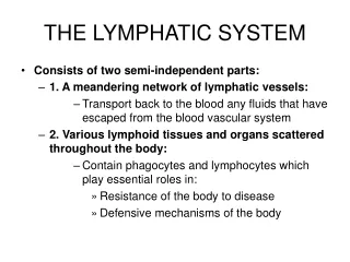
20. The Lymphatic System. Lymphatic System: Overview. Consists of two semi-independent parts: A network of lymphatic vessels Lymphoid tissues and organs scattered throughout the body Returns interstitial fluid and leaked plasma proteins back to the blood

E N D
20 The Lymphatic System
Lymphatic System: Overview • Consists of two semi-independent parts: • A network of lymphatic vessels • Lymphoid tissues and organs scattered throughout the body • Returns interstitial fluid and leaked plasma proteins back to the blood • Lymph – interstitial fluid once it has entered lymphatic vessels
Lymphatic System: Overview Figure 20.2a
Lymphatic System: Overview Figure 20.1a
Lymphatic Vessels • One-way system, lymph flows toward the heart • Lymph vessels include: • Microscopic, permeable, blind-ended capillaries • Lymphatic collecting vessels • Trunks and ducts
Lymphatic Capillaries • Similar to blood capillaries, with modifications: • Very permeable • Loosely joined endothelial minivalves • Withstand interstitial pressure and remain open • The minivalves function as one-way gates that: • Allow interstitial fluid to enter lymph capillaries • Do not allow lymph to escape from the capillaries
Lymphatic Capillaries Figure 20.1b
Lymphatic Capillaries • During inflammation, lymph capillaries can absorb: • Cell debris • Pathogens • Cancer cells • Cells in the lymph nodes cleanse and “examine” this debris • Lacteals – specialized lymph capillaries present in intestinal mucosa • Absorb digested fat and deliver chyle to the blood
Lymphatic Collecting Vessels • Have the same three tunics as veins • Have thinner walls, with more internal valves • Anastomose more frequently • Collecting vessels in the skin travel with superficial veins • Deep vessels travel with arteries • Nutrients are supplied from branching vasa vasorum
Lymphatic Trunks • Lymphatic trunks are formed by the union of the largest collecting ducts • Major trunks include: • Paired lumbar, bronchomediastinal, subclavian, and jugular trunks • A single intestinal trunk
Lymphatic Trunks • Lymph is delivered into one of two large trunks • Right lymphatic duct – drains the right upper arm and the right side of the head and thorax • Thoracic duct – arises from the cisterna chyli and drains the rest of the body
Lymphatic Trunks Figure 20.2b
Lymph Transport • The lymphatic system lacks a pumping organ • Vessels are low-pressure conduits • Uses the same methods as veins to propel lymph: • Pulsations of nearby arteries • Contractions of smooth muscle in the walls of the lymphatics
Lymphoid Cells • Lymphocytes are the main cells involved in the immune response • Two main varieties: • T cells • B cells
Lymphocytes • T cells and B cells protect the body against antigens • Antigen – anything the body perceives as foreign • Bacteria and their toxins; viruses • Mismatched RBCs or cancer cells
Lymphocytes • T cells • Manage the immune response • Attack and destroy foreign cells • B cells • Produce plasma cells, which secrete antibodies • Antibodies immobilize antigens
Other Lymphoid Cells • Macrophages – phagocytize foreign substances and help activate T cells • Dendritic cells – spiny-looking cells with functions similar to macrophages • Reticular cells – fibroblast–like cells that produce a stroma, or network, that supports other cell types in lymphoid organs
Lymphoid Tissue • Diffuse lymphatic tissue – scattered reticular tissue elements in every body organ • Larger collections appear in the lamina propria of mucous membranes and lymphoid organs • Lymphatic follicles (nodules) – solid, spherical bodies consisting of tightly packed reticular elements and cells • Germinal center composed of dendritic and B cells • Found in isolation and as part of larger lymphoid organs
Lymph Nodes • Principal lymphoid organs of the body • Embedded in connective tissue and clustered along lymphatic vessels • Aggregations of these nodes occur near the body surface in inguinal, axillary, and cervical regions of the body Figure 20.4a
Lymph Nodes • Two basic functions: • Filtration – macrophages destroy microorganisms and debris • Immune system activation – monitor for antigens and mount an attack against them
Structure of a Lymph Node • Nodes are bean shaped and surrounded by a fibrous capsule • Trabeculae extended inward from the capsule and divide the node into compartments • Nodes have two histologically distinct regions: a cortex and a medulla Figure 20.4a
Structure of a Lymph Node • Cortex contains follicles with germinal centers, heavy with dividing B cells • Dendritic cells nearly encapsulate the follicles • Deep cortex houses T cells in transit • T cells circulate continuously among the blood, lymph nodes, and lymphatic stream
Structure of a Lymph Node • Medullary cords extend from the cortex and contain B cells, T cells, and plasma cells • Throughout the node are lymph sinuses crisscrossed by reticular fibers • Macrophages reside on these fibers and phagocytize foreign matter
Structure of a Lymph Node Figure 20.4a, b
Circulation in the Lymph Nodes • Lymph enters via afferent lymphatic vessels • It then enters a large subcapsular sinus and travels into smaller sinuses • It meanders through these sinuses and exits the node at the hilus via efferent vessels • Because there are fewer efferent vessels, lymph stagnates somewhat in the node • This allows lymphocytes and macrophages time to carry out protective functions
Lymphoid Organs Figure 20.5
Other Lymphoid Organs • The spleen, thymus gland, and tonsils • Peyer’s patches and bits of lymphatic tissue scattered in connective tissue • All are composed of reticular connective tissue • All help protect the body • Only lymph nodes filter lymph
Spleen • Largest lymphoid organ, located on the left side of the abdominal cavity beneath the diaphragm • It is served by the splenic artery and vein, which enter and exit at the hilus • Functions: • Site of lymphocyte proliferation • Immune surveillance and response • Cleanses the blood
Additional Spleen Functions • Stores breakdown products of RBCs for later reuse • Spleen macrophages salvage and store iron for later use by bone marrow • Site of fetal erythrocyte production (normally ceases after birth) • Stores blood platelets
Structure of the Spleen • Surrounded by a fibrous capsule, it has trabeculae that extend inward and contains lymphocytes, macrophages, and huge numbers of erythrocytes • Two distinct areas: • White pulp – containing mostly lymphocytes suspended on reticular fibers and involved in immune functions • Red pulp – remaining splenic tissue concerned with disposing of worn-out RBCs and bloodborne pathogens
Structure of the Spleen Figure 20.6a, b
Thymus • A bilobed organ that secretes hormones (thymosin and thymopoietin) that cause T lymphocytes to become immunocompetent • Size of the thymus varies with age: • In infants, it is found in the inferior neck and extends into the mediastinum where it partially overlies the heart • It increases in size and is most active during childhood • It stops growing during adolescence and then gradually atrophies
Internal Anatomy of the Thymus • Thymic lobes contain an outer cortex and inner medulla • Cortex contains densely packed lymphocytes and scattered macrophages • Medulla contains fewer lymphocytes and thymic (Hassall’s) corpuscles
Thymus • The thymus differs from other lymphoid organs in important ways • It functions strictly in T lymphocyte maturation • It does not directly fight antigens • The stroma of the thymus consists of star-shaped epithelial cells (not reticular fibers) • These thymocytes secrete the hormones that stimulate lymphocytes to become immunocompetent
Tonsils • Simplest lymphoid organs; form a ring of lymphatic tissue around the pharynx • Location: • Palatine tonsils – either side of the posterior end of the oral cavity • Lingual tonsils – lie at the base of the tongue • Pharyngeal tonsil – posterior wall of the nasopharynx • Tubal tonsils – surround the openings of the auditory tubes into the pharynx
Tonsils • Lymphoid tissue of tonsils contains follicles with germinal centers • Tonsil masses are not fully encapsulated • Epithelial tissue overlying tonsil masses invaginates, forming blind-ended crypts • Crypts trap and destroy bacteria and particulate matter
Aggregates of Lymphoid Follicles • Peyer’s patches – isolated clusters of lymphoid tissue, similar to tonsils • Found in the wall of the distal portion of the small intestine • Similar structures are found in the appendix • Peyer’s patches and the appendix: • Destroy bacteria, preventing them from breaching the intestinal wall • Generate “memory” lymphocytes for long-term immunity
MALT • MALT – mucosa-associated lymphatic tissue: • Peyer’s patches, tonsils, and the appendix (digestive tract) • Lymphoid nodules in the walls of the bronchi (respiratory tract) • MALT protects the digestive and respiratory systems from foreign matter
Developmental Aspects • Beginnings of the lymphatic vessels and main clusters of lymph nodes are apparent by the fifth week of embryonic development • These arise from the budding of lymph sacs from developing veins • Lymphatic organs (except the thymus) arise from mesoderm
Developmental Aspects • The thymus (endodermal origin) forms as an outgrowth of the pharynx • Except for the spleen and tonsils, lymphoid organs are poorly developed at birth