Download
1 / 56
610 likes | 924 Vues
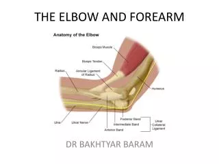
Chapter 19: The Elbow, Forearm, Wrist and Hand. Anatomy of the Elbow. Assessment of the Elbow. History Past history Mechanism of injury When and where does it hurt? Motions that increase or decrease pain Type of, quality of, duration of, pain? Sounds or feelings?

E N D
Assessment of the Elbow • History • Past history • Mechanism of injury • When and where does it hurt? • Motions that increase or decrease pain • Type of, quality of, duration of, pain? • Sounds or feelings? • How long were you disabled? • Swelling? • Previous treatments?
Assessment (cont’d.) • Observations • Deformities and swelling? • Carrying angle • Cubitus valgus versus cubitus varus • Flexion and extension • Cubitus recurvatum • Elbow hyperextension? • Palpation • Be sure to check sites of pain and deformity • Assess epicondyles, olecranon, distal aspect of humerus and proximal aspect of ulna • Soft tissue – muscles, tendons, joint capsules and ligaments surrounding joint
Prevention of Elbow, Forearm and Wrist Injuries • Vulnerable to a variety of acute and chronic injuries • Protective gear is always recommended to reduce severity of injury • Chronic injury reduction • Limit repetitions (baseball, tennis) • Utilize proper mechanics • Use equipment that is appropriate for skill level • Maintain appropriate levels of strength, flexibility, and endurance for activity
Olecranon Bursitis • Cause of Injury • Superficial location makes it extremely susceptible to injury (acute or chronic) --direct blow • Signs of Injury • Pain, swelling, and point tenderness • Swelling will appear almost spontaneously and w/out usual pain and heat
Olecranon Bursitis (cont’d.) Care In acute conditions, ice Chronic cases require protective therapy If swelling fails to resolve, aspiration may be necessary Can be padded in order to return to competition
Elbow Sprain • Cause of Injury • Elbow hyperextension or a valgus force (often seen in the cocking phase of throwing • Signs of Injury • Pain along medial aspect of elbow • Inability to grasp objects • Point tenderness over the MCL • Care • Conservative treatment begins w/ RICE elbow fixed at 90 degrees in a sling for at least 24 hours • Gradual work on elbow ROM • Athlete should modify activity • Gradual progression involving an increase in number of throws while range and strength return • If unstable, MCL can be reconstructed • Tommy John’s procedure
Lateral Epicondylitis • AKA - Tennis Elbow • Cause of Injury • Repetitive microtrauma to insertion of extensor muscles of lateral epicondyle • Signs of Injury • Aching pain in region of lateral epicondyle after activity • Pain worsens and weakness in wrist and hand develop • Elbow has decreased ROM; pain w/ resistive wrist extension
Lateral Epicondylitis (cont’d.) • Care • RICE, NSAID’s and analgesics • ROM exercises and PRE, deep friction massage, hand grasping while in supination, avoidance of pronation motions • Mobilization and stretching in pain free ranges • Use of a counter force or neoprene sleeve • Proper mechanics and equipment instruction is critically important
Medial Epicondylitis • Cause of Injury • Repeated forceful flexion of wrist and extreme valgus torque of elbow • Signs of Injury • Pain produced w/ forceful flexion or extension • Point tenderness and mild swelling • Passive movement of wrist seldom elicits pain, but active movement does • Care • Sling, rest, cryotherapy or heat through ultrasound • Analgesic and NSAID's • Curvilinear brace below elbow to reduce elbow stressing • Severe cases may require splinting and complete rest for 7-10 days
Osteochondritis Dessicans • Cause of Injury • Impairment of blood supply to anterior surface resulting in degeneration of articular cartilage, and bone creating loose bodies within the joint • Signs of Injury • Sudden pain, locking; range usually returns in a few days • Swelling, pain and crepitation may also occur • Care • If repeated locking occurs, loose bodies may be removed surgically • Without removal, arthritis may develop
Ulnar Nerve Injuries • Cause of Injury • Pronounced cubital valgus may cause deep friction problem • Ulnar nerve dislocation • Traction injury from valgus force, irregularities w/ tunnel, subluxation of ulnar nerve due to lax impingement, or progressive compression of ligament on the nerve • Signs of Injury • Generally respond with paresthesia in 4th and 5th fingers • Care • Conservative management – avoid aggravating condition • Surgery may be necessary if stress on nerve can not be avoided
Elbow Dislocation • Cause of Injury • High incidence in sports caused by fall on outstretched hand w/ elbow extended or severe twist while flexed • Signs of Injury • Swelling, severe pain, disability • May be displaced backwards, forward, or laterally • Complications w/ median and radial nerves and blood vessels • Rupture and tearing of stabilizing ligaments will usually accompany the injury • Care • Immobilize and refer to physician for reduction • Following reduction, elbow should remain splinted in flexion for 3 weeks
Elbow Fractures • Cause of Injury • Fall on flexed elbow or from a direct blow • Fracture can occur in any one or more of the bones • Fall on outstretched hand often fractures humerus above condyles or between condyles • Signs of Injury • May or may not result in visual deformity • Hemorrhaging, swelling, muscle spasm • Care • Ice and sling for support – refer to physician
Assessment of the Forearm • History • What was the cause? • What were the symptoms at the time of injury, did they occur later, were they localized or diffuse? • Was there swelling an discoloration? • What treatment was given and how does it feel now? • When did the injury occur?
Assessment (cont’d.) • Observation • Visually inspect for deformities, swelling and skin defects • Range of motion • Pain w/ motion • Palpation • Palpated at distant sites and at point of injury • Can reveal tenderness, edema, fracture, deformity, changes in skin temperature, a false joint, bone fragments or lack of bone continuity
Forearm Contusions • Cause of Injury • Ulnar side receives majority of blows due to arm blocks • Can be acute or chronic • Result of direct contact or blow • Signs of Injury • Pain, swelling and hematoma • If repeated blows occur, heavy fibrosis and possibly bony callus could form w/in hematoma
Forearm Contusions (cont’d.) • Care • Proper care in acute stage involves RICE for at least one hour and followed up w/ additional cryotherapy • Protection is critical - full-length sponge rubber pad can be used to provide protective covering
Forearm Strains • Cause of Injury • Forearm strain - most come from severe static contraction • Signs of Injury • Dull ache between extensors which cross posterior aspect of forearm • Weakness and pain w/ contraction • Point tenderness in interosseus membrane • Care • Treat symptomatically • If occurs early in season, strengthen forearm; when it occurs late in season treat w/ cryotherapy, wraps, or heat
Forearm Fractures • Cause of Injury • Common in youth - due to falls and direct blows • Fracturing ulna or radius singularly is rarer than simultaneous fractures to both • Signs of Injury • Audible pop or crack followed by moderate to severe pain, swelling, and disability • Edema, ecchymosis w/ possible crepitus • Older athlete may experience extensive damage to soft tissue structures
Forearm Fractures • Care • RICE, splint, immobilize and refer to physician • Athlete is usually incapacitated for 8 weeks
Colles’ Fracture • Cause of Injury • Occurs in lower end of radius and ulna • MOI is fall on extended wrist, forcing radius and ulna into hyperextension • A Smith fracture involves falling on flexed wrist • Less common
Colles’ Fracture (cont’d.) • Signs of Injury • Forward displacement of radius causing visible deformity (silver fork deformity) • When no deformity is present, injury may be passed off as bad sprain • Extensive bleeding and swelling • Tendons may be torn/avulsed and there may be median nerve damage • Care • Cold compress, splint wrist and refer to physician • X-ray and immobilization • Without complications a Colles’ fracture will keep an athlete out for 1-2 months
Assessment of the Wrist, Hand and Fingers • History • Past history • Mechanism of injury • When does it hurt? • Type of, quality of, duration of, pain? • Sounds or feelings? • How long were you disabled? • Swelling? • Previous treatments?
Assessment (cont’d.) • Observation • Postural deviations • Is the part held still, stiff or protected? • Wrist or hand swollen or discolored? • General attitude • What movements can be performed fully and rhythmically? • Thumb to finger touching • Color of nailbeds
Assessment (cont’d.) • Palpation • Begin with palpation of bony structures • Palpate for pain and deformity • Be sure to palpate all the bones of wrist and hand during the evaluation • Soft tissue palpation should include the tendons crossing the wrist and the muscles involved in movement of the thumb as well as the digits
Wrist Sprains • Cause of Injury • Most common wrist injury • Arises from any abnormal, forced movement • Falling on hyperextended wrist, violent flexion or torsion • Signs of Injury • Pain, swelling and difficulty w/ movement
Wrist Sprains (cont’d.) • Care • Refer to physician for X-ray if severe • RICE, splint and analgesics • Have athlete begin strengthening soon after injury • Tape for support can benefit healing and prevent further injury
Wrist Tendinitis • Cause of Injury • Primary cause is overuse of the wrist • Repetitive wrist accelerations and decelerations • Signs of Injury • Pain on active use or passive stretching • Tenderness and swelling over involved tendon • Care • Acute pain and inflammation treated w/ ice massage 4x daily for first 48-72 hours, NSAID’s and rest • Use of wrist splint may protect injured tendon • PRE can be instituted once swelling and pain subsided (high rep, low resistance)
Carpal Tunnel Syndrome • Cause of Injury • Compression of median nerve due to inflammation of tendons and sheaths of carpal tunnel • Result of repeated wrist flexion or direct trauma to anterior aspect of wrist • Signs of Injury • Sensory and motor deficits (tingling, numbness and paresthesia); weakness in thumb • Care • Conservative treatment - rest, immobilization, NSAID’s • If symptoms persist, corticosteroid injection may be necessary or surgical decompression of transverse carpal ligament
Scaphoid Fracture • Cause of Injury • Caused by force on outstretched hand, compressing scaphoid between radius and second row of carpal bones • Signs of Injury • Swelling, severe pain in anatomical snuff box • Care • Must be splinted and referred for X-ray prior to casting • May be missed on initial X-ray • Immobilization lasts 6 weeks and is followed by strengthening and protective tape • Wrist requires protection against impact loading for 3 additional months • Often fails to heal due to poor blood supply
Hamate Fracture • Cause of Injury • Occurs as a result of a fall or more commonly from contact while athlete is holding an implement • Signs of Injury • Wrist pain and weakness (5th digit due to ulnar nerve compression), along w/ point tenderness • Care • Casting wrist and thumb is treatment of choice • Hook of hamate can be protected w/ doughnut pad to take pressure off area
Ganglion Cyst • Cause of Injury • Synovial cyst (herniation of joint capsule or synovial sheath of tendon) • Generally appears following wrist strain or repeated forced hyperextension • Signs of Injury • Appear on back of wrist generally • Occasional pain w/ lump at site • Pain increases w/ use • May feel soft, rubbery or very hard
Ganglion Cyst (cont’d.) • Care • Old method was to first break down the swelling through distal pressure and then apply pressure pad to encourage healing • New approach includes aspiration, chemical cauterization w/ subsequent pressure from pad • Surgical removal is most effective way
Metacarpal Fracture • Cause of Injury • Direct axial force or compressive force • Fractures of the 5th metacarpal are associated w/ boxing or martial arts (boxer’s fracture) • Signs of Injury • Pain and swelling; possible angular or rotational deformity • Palpable defect is possible • When patient makes a fist the knuckle will be depressed or sunken • Care • RICE, refer to physician for reduction and immobilization • Deformity is reduced, followed by splinting - 4 weeks
Mallet Finger • Cause of Injury • Caused by a blow that contacts tip of finger avulsing extensor tendon from insertion • Signs of Injury • Pain at DIP; X-ray shows avulsed bone on dorsal proximal distal phalanx • Unable to extend distal end of finger (carrying at 30 degree angle) • Point tenderness at sight of injury • Care • RICE and splinting (in extension) for 6-8 weeks
Boutonniere Deformity • Cause of Injury • Rupture of extensor tendon dorsal to the middle phalanxForces DIP joint into extension and PIP into flexion • Signs of Injury • Severe pain, obvious deformity and inability to extend DIP joint • Swelling, point tenderness • Care • Cold application, followed by splinting of PIP • Splinting must be continued for 5-8 weeks • Athlete is encouraged to flex distal phalanx
Jersey Finger • Cause of Injury • Rupture of flexor digitorum profundus tendon from insertion on distal phalanx • Often occurs w/ ring finger when athlete tries to grab a jersey • Signs of Injury • DIP can not be flexed, finger remains extended • Pain and point tenderness over distal phalanx • Care • Must be surgically repaired • Rehab requires 12 weeks and there is often poor gliding of tendon, w/ possibility of re-rupture