Download
1 / 43
430 likes | 437 Vues
The Disease of Hematopoietic and Lymphoid Systems. Hematopoietic and Lymphoid Systems. Myeloid tissue Bone marrow RBC, platelets, granulocytes, monocytes Lymphoid tissue lymph nodes thymus spleen. Disorders encompass a wide range of diseases, involving its any organs or tissues.

E N D
The Disease of Hematopoietic and Lymphoid Systems
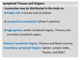
Hematopoietic and Lymphoid Systems • Myeloid tissue • Bone marrow • RBC, platelets, granulocytes, monocytes • Lymphoid tissue • lymph nodes • thymus • spleen
Disorders • encompass a wide range of diseases, involving its any organs or tissues. • RBC: usually anemia • WBC: overgrowth, usually malignant common and lethal
Neoplastic proliferations of white cells Be defined briefly as follows: • Myeloid neoplasms髓样组织肿瘤 • Lymphoid neoplasms 淋巴样组织肿瘤 • Histiocytosis Man-made classification The distinction in some cases may be blurred.
Myeloid neoplasms • Arise within hematopoietic stem cells • Three general categories: • Acute myelogenous leukemia (AML 急性髓性白血病) • Chronic myeloproliferative disorders(慢性骨髓增生性疾病) • Chronic myelogenous leukemia 慢性髓性白血病 • Polycythemia vera 真性红细胞增生症 • Myeloid metaplasia with myelofibrosis 伴骨髓纤维化的髓样增生 • Essential thrombocythemia 真性血小板增生症 • Myelodysplastic syndromes(骨髓增生异常综合症) 8 types: FAB classification M1~8 Myeloid neoplasms: CD13,CD14,CD15,CD64
Lymphoid neoplasms • A group of entities that vary widely in terms of their clinical presentation and behavior • Classification scheme that relies on a combination of clinical, morphologic, phenotypic and genotypic features the relationship between lymphoma and lymphocytic leukemia • Certain important relevant principles most: B Cell origin neoplasms are often composed of cells that arrest at specific stages
Lymphoid neoplasms ⅠA. Precursor B-cell neoplasms ⅠB. Peripheral B-cell neoplasms ⅡA. Precursor T-cell neoplasms ⅡB. Peripheral T-/NK-cell neoplasms Ⅲ. Hodgkin lymphoma NHL
ⅠA. Precursor B-cell neoplasms 1. Precursor B-cell leukemia/lymphoma ⅡA. Precursor T-cell neoplasms 1. Precursor T-cell leukemia/lymphoma pre-B-lymphoblastic tumor: first leukemia lymphoma pre-T-lymphoblastic tumor: mediastinal masses involved the thymus leukemia Both tumors usually have the clinical appearance of an acute lymphoblastic leukemia (ALL)
Common features of acute leukemias ALL +AML • Clinical features 1.Abrupt stormy onset—within 3 months 2. Symptoms related to depression of normal marrow function Pathophysiology Block in differentiation blasts accumulate in marrow normal hematopoietic stem cells Anemia fatigue Thromobocytopenia bleeding Mature leukocytes infection
Common features of acute leukemias • Clinical features 3. Bone pain and tenderness 4. Generalized lymphadenopathy splenomegaly, and hepatomegaly More common in ALL 5.Central nervous system manifestations Headache, vomiting, and nerve palsies children >adults, ALL>AML
Common features of acute leukemias • Laboratory findings • Anemia • Platelet count is usually depressed • WBC count is variably elevated Much more important: blasts in the circulating blood and the bone marrow
Common features of acute leukemias • Clinical treatment Chemotherapy Bone marrow stem cell(MSCs) transplantation
AML ALL Different features between AML and ALL Pre-B(85% ): children, 4 years; Pre-T(15%): adolescent males, 15-20years Age adults; median age : 50 yrs Morphology Fine chromatin; 3-5 nucleoli, more cytoplasm containing granules, Auer rods coarse and clumped chromatin 1-2 nucleoli histochemistry Peroxidase + PAS+
AML ALL Clinical features immunophenotyping TdT (95% +) B: CD19 T: CD2 As described previously CD13,CD14,CD15, CD64 M2: t(8;21) M3:t(15;17) 维甲酸治疗 M4: inv(16) /del(16) Karyotypic changes Pre-B: Hyperdiploidy t (12;21) ;Ph Good:2-10 yrs, pre-B prognosis Devastating disease
Chronic myelogenous leukemia, CML Age 25-60 years Peak incidence : 40-50 years Clinical features 1. Onset is slow; 2. nonspecific initial symptoms Easy fatigability, weakness, and weight loss 3. Extreme splenomegaly infarct pains
Chronic myelogenous leukemia, CML Pathology changes Leukocyte count↑↑and bone marrow is hypercellular Predominantly: neutrophils and myelocytes Basophils and eosinophils myeloblasts<10% Spleen infarcts acute onset left upper quadrant pain
Chronic myelogenous leukemia, CML • Karyotypic changes • Ph (Philadelphia) chromosome: t(9;22)(q34;q11) • c-abl-ber fusion genes/P210protein: • increased, dysregulated tyrosine kinase activity • Prognosis • Slow progression • without treatment, median survival is 3 years • Bone marrow transplantation • STI-571: the exciting concept of designer drugs that • specifically target oncoproteins
ⅠB. Peripheral B-cell neoplasms • Small lymphocytic lymphoma/chronic lymphocytic leukimia(慢性淋巴细胞性白血病/小淋巴细胞性淋巴瘤) • B-cell prolymphocytec leukemia B细胞性前淋巴细胞白血病 • Lymphoplasmacytic lymphoma 淋巴细胞、桨细胞性淋巴瘤 • Mantle cell lymphoma 套细胞性淋巴瘤 • Follicular Lymphoma 滤泡性淋巴瘤 • Extranodal marginal zone lymphoma(MALT lymphoma) 节外边缘带淋巴瘤 7. Splenic marginal zone lymphoma 脾边缘带淋巴瘤 8. Nodal marginal zone lymphoma 淋巴结边缘带淋巴瘤 9. Hairy cell leukemia 毛细胞性白血病 10.Plasmacytoma/plasma cell myeloma 浆细胞瘤/骨髓瘤 11. Diffuse Large B-cell Lymphoma 弥漫性大细胞性淋巴瘤 12. Burkitt Lymphoma 伯基特淋巴瘤
Peripheral Lymphoid Cell Tumors • Age • >50 years • Clinical features • 1.Often asymptomatic or nonspecific • (easy fatigability, weight loss, and anorexia) • 2. Hypogammaglobulinemia • susceptibility to bacterial infections • 3. Generalized lymphadenopathy, splenohepatomegaly • 4. Total leukocyte count Small lymphocytic lymphoma/ chronic lymphocytic leukimia
Small lymphocytic lymphoma/chronic lymphocytic leukimia Lymph nodes, the bone marrow, spleen, liver Morphology Low power: sheets of tumor cells diffusely efface involved lymph nodes
Small lymphocytic lymphoma/chronic lymphocytic leukimia Morphology Predominant cells compact, small, dark-staining round nuclei, Scanty cytoplasm, and little variation in size Proliferation centers--Pseudofollicle foci of mitotically active prolymphocytes
Small lymphocytic lymphoma/chronic lymphocytic leukimia Immunophenotyping: mature Bcells Pan-B-cell markers:CD19,CD20, SIg Prognosis the course and prognosis are extremely variable The median survival is 4~6years. To transform to more aggressive tumors, such as diffuse large B-cell lymphoma: less than 1 year
Follicular Lymphoma Age Older persons (rarely before age 20 years) Clinical characteristics Painless lymphadenopathy, frequently generalized Morphology lymph nodes are effaced by a nodular or follicular architecture
Follicular Lymphoma Morphology Predominant neoplastic cells---centrocyte, CC 1. slight larger than resting lymphocytes 2. Coarse and condensed chromatin prominent indentations and linear infoldings Nucleoli are indistinct Other cells ---Centroblast,CB 1.3-4 times the size of resting lymphocytes 2. Vesicular chromatin several nucleoli modest amounts of cytoplasm
Immunophenotyping Pan-B-cell markers: CD19, CD20, BCL2 BCL2 14: IgH gene; 18:BCL2 gene Translocation t(14,18) Bcl-2 protein antiapoptotic effect
Prognosis Indolent course, median survival,7 to 9 years not easily curable 40% of patients progress to a diffuse large B-cell lymphoma
diffuse large B-cell lymphoma Age Most important type of lymphoma in adults approximatelly 50% of all adult NHLs Morphology its nuclei is large and varible (3-4 times the size of resting lymphocytes)
diffuse large B-cell lymphoma Immunophenotype Pan-B-cell marker: CD19, CD20 Subtypes: epstein-Barr virus(EBV) associated acquired immunodeficiency syndrome and iatrogenic immunosuppression human herpes type 8( HHV-8) infections a rare group mediastinal large B-cell lymphoma usually in young adults a predilection for spread to abdominal viscera and CNS
Karyotype 30% t(14;18) BCL2 arrangement 1/3 3q27 BCL6 arrangement Prognosis Aggressive , Rapidly fatal if untreated With intensive combination chemotherapy, complete remission can be achieved in 60%-80% of the patients
Burkitt Lymphoma • Age • Children or young adults • endemic in some parts of Africa and sporadic in other areas. • Common sites • Africa : Maxilla(上颌骨) or mandible (下颚骨) • other areas: abdominal (bowel, retroperitoneum, ovaries) • Pathogenesis • Correlated with EBV infection
Morphology • Starry sky: macrophages with ingested nuclear debris • Monotonous • round or oval nuclei containing 2-5 prominent nucleoli • Active mitosis
Burkitt Lymphoma Immunophenotype SIgM, pan-B-cell markers: CD19, CD10 Karyotype t(8;14) et al Translocations involving MYC gene 8 MYC 14 IgH or 2、22 К,λ Prognosis It may be the fastest growing human neoplasm, however, with very aggressive chemotherapy, the majority of patients can be cured.
Ⅲ. Hodgkin Lymphoma 10%~20%, Tomas Hodgkin It was separated from NHLs for several reasons • It arises almost invariably in a single node or a chain of nodes • distinctive neoplastic giant cells called Read-Sternberg (RS) cells mixed with a variable infiltrate of reactive inflammatory cells. • Systemic manifestations, such as fever • With a relatively good prognosis.
Ⅲ. Hodgkin Lymphoma RS cells Abundant cytoplasm, d=15 to 45um Multilobate nucleus or multinucleate with large, round, prominent nucleoli Mirror-image nuclei
Ⅲ. Hodgkin Lymphoma Variant of RS cells • Mononuclear variants • Lacunar cell A large folded or hyperlobate nucleus with multiple small nucleoli Abundant, pale-staining cytoplasm
Ⅲ. Hodgkin Lymphoma Variant of RS cells 3.Lymphocytic and histocytic variants,(L&H cell or Popcorn cell ) have twisted , multilobate nuclei, resembling popcorn kernels
Hodgkin lymphoma 1. Nodular lymphocyte predominant Hodgkin lymphoma CD45 +; CD15, CD30 – 2. Classical Hodgkin lymphoma CD45 -; CD15, CD30 +
Nodular lymphocyte predominant Hodgkin lymphoma Morphology L&H cells: nodular or diffuse Many small lymphocytes, histocytes, epitheloid cells: nodular or diffuse Immunophenotype CD20, CD79, BCL6, and CD45 + CD15, CD30 – Young male; <35yrs Good prognosis
Classical Hodgkin lymphoma • Nodular sclerosis • Mixed cellularity • Lymphocyte rich • Lymphocyte depleted
Classical Hodgkin lymphoma 1. Nodular sclerosis Hodgkin lymphoma The most common, young female Morphology Collagen bands divide the lymphoid tissue into circumscribed nodules Lacunar cell Lymphocytes, eosinophils, Histiocytes Prognosis is good.
Classical Hodgkin lymphoma 2. Mixed cellularity Hodgkin lymphoma More common in older male Typical RS and mononuclear cells are plentiful Lymphocytes, eosinophils, plasma cells, histiocytes
Classical Hodgkin lymphoma 3. Lymphocyte predominance young male A large number of small, mature-looking reactive lymphocytes admixed with a variable number of benign histiocytes, often within large, poorly defined nodules L&H is common and typical RS is rare
Classical Hodgkin lymphoma 4. Lymphocyte depletion Vary rare RS cells: diffusely proliferate Lymphocytes: greatly reduce Old people, HIV infection Poor prognosis