Download
1 / 44
440 likes | 482 Vues
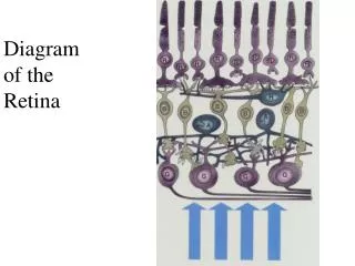
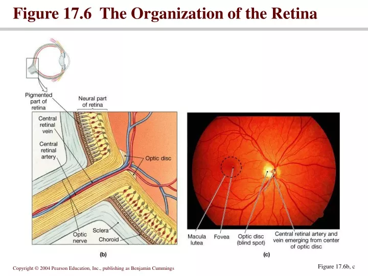
Figure 17.6 The Organization of the Retina. Figure 17.6b, c. Eye anatomy. Ciliary body and lens divide the anterior cavity of the eye into posterior (vitreous) cavity and anterior cavity Anterior cavity further divided anterior chamber in front of eye

E N D
Figure 17.6 The Organization of the Retina Figure 17.6b, c
Eye anatomy • Ciliary body and lens divide the anterior cavity of the eye into posterior (vitreous) cavity and anterior cavity • Anterior cavity further divided • anterior chamber in front of eye • posterior chamber between the iris and the lens
Figure 17.8 The Circulation of Aqueous Humor Figure 17.8
Fluids in the eye • Aqueous humor circulates within the eye • diffuses through the walls of anterior chamber • passes through canal of Schlemm • re-enters circulation • Vitreous humor fills the posterior cavity. • Not recycled – permanent fluid
Lens • Posterior to the cornea and forms anterior boundary of posterior cavity • Posterior cavity contains vitreous humor • Lens helps focus • Light is refracted as it passes through lens • Accommodation is the process by which the lens adjusts to focus images • Normal visual acuity is 20/20
Figure 17.9 Image Formation Figure 17.9
Figure 17.10 Accommodation Figure 17.10
Figure 17.11 Visual Abnormalities Figure 17.11
Visual physiology • Rods – respond to almost any photon • Cones – specific ranges of specificity
Figure 17.13 Rods and Cones Figure 17.13
Photoreceptor structure • Outer segment with membranous discs • Narrow stalk connecting outer segment to inner segment • Light absorption occurs in the visual pigments • Derivatives of rhodopsin
Figure 17.14 Photoreception Animation: Photoreception PLAY Figure 17.14a, b
Figure 17.14 Photoreception Figure 17.14c, d
Figure 17.15 Bleaching and Regeneration of Visual Pigments Figure 17.15
Color sensitivity • Integration of information from red, blue and green cones • Colorblindness is the inability to detect certain colors
retinal adaptation • Dark adapted – most visual pigments are fully receptive to stimulation • Light adapted – pupil constricts and pigments bleached.
the visual pathway • Large M-cells monitor rods • Smaller more numerous P cells monitor cones
Figure 17.18 Convergence and Ganglion Cell Function Figure 17.18
Seeing in stereo • Vision from the field of view transfers from one side to the other while in transit • Depth perception is obtained by comparing relative positions of objects from the two eyes
Figure 17.19 The Visual Pathways Figure 17.19
Visual circadian rhythm • Input to suprachiasmic nucleus affects the function of the brainstem • Circadian rhythm ties to day-night cycle, and affects metabolic rates
Both equilibrium and hearing are provided by receptors of the inner ear Anatomy of the ear – External Ear • Auricle or pinnae surrounds the ear • External acoustic meatus ends on tympanic membrane
Figure 17.20 The Anatomy of the Ear Figure 17.20
Middle ear • Communicates with pharynx via pharyngotympanic membrane • Middle ear encloses and protects the auditory ossicles
Figure 17.21 The Middle Ear Figure 17.21
Inner ear • Membranous labyrinth contains endolymph • Bony labyrinth surrounds and protects membranous labyrinth • Vestibule • Semicircular canals • Cochlea
Figure 17.22 The Inner Ear Figure 17.22
Components of the inner ear • Vestibule contains the utricle and saccule • Semicircular canals contain the semicircular ducts • Cochlea contains the cochlear duct
Windows • Round window separates the perilymph from the air spaces of the middle ear • Oval window connected to the base of the stapes • Basic receptors of inner ear are hair cells • Provide information about the direction and strength of stimuli
Equilibrium • Anterior, posterior and lateral semicircular ducts are continuous with the utricle • Each duct contains an ampulla with a gelatinous cupula and associated sensory receptor • Saccule and utricle connected by a passageway continuous with the endolymphatic duct • Terminates in the endolymphatic sac • Saccule and utricle have hair cells clustered in maculae • Cilia contact the otolith (statoconia)
Figure 17.23 The Vestibular Complex Figure 17.23a, b, & d
Figure 17.23 The Vestibular Complex Figure 17.23c
Figure 17.23 The Vestibular Complex Figure 17.23e
Vestibular neural pathway • Vestibular receptors activate sensory neurons of the vestibular ganglia • Axons form the vestibular branch of cranial nerve VII • Synapses within the vestibular nuclei
Figure 17.24 Pathways for Equilibrium Sensation Figure 17.24
Hearing • Cochlear duct lies between the vestibular duct and the tympanic duct • Hair cells of the cochlear duct lie within the Organ of Corti • Intensity is the energy content of a sound • Measured in decibels
Figure 17.25 The Cochlea Figure 17.25a, b
Figure 17.26 The Organ Of Corti Figure 17.26a, b
Pathway of sound • Sound waves travel toward tympanic membrane, which vibrates • Auditory ossicles conduct the vibration into the inner ear • Tensor tympani and stapedius muscles contract to reduce the amount of movement when loud sounds arrive • Movement at the oval window applies pressure to the perilymph of the cochlear duct • Pressure waves distort basilar membrane • Hair cells of the Organ of Corti are pushed against the tectoral membrane
Figure 17.28 Sound and Hearing Figure 17.28a
Figure 17.29 Sound and Hearing Figure 17.29
Neural pathway • Sensory neurons of hearing are located in the spiral ganglion of the cochlea • Afferent fibers form the cochlear branch of cranial nerve VIII • Synapse at the cochlear nucleus
You should now be familiar with: • The sensory organs of smell, and the olfactory pathways in the brain. • The accessory and internal structures of the eye, and their functions. • How light stimulates the production of nerve impulses, and the visual pathways. • The structures of the external and middle ear and how they function. • The parts of the inner ear and their roles in equilibrium and hearing. • The pathways for the sensations of equilibrium and hearing.