Download
1 / 36
360 likes | 382 Vues
Learn the importance of capnography in respiratory and ventilation monitoring, tube placement verification, and assessing conditions like asthma, head injuries, and cardiac arrest.

E N D
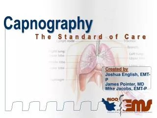
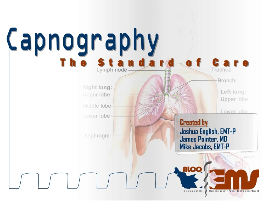
Capnography The Standard of Care Created by Joshua English, EMT-P James Pointer, MD Mike Jacobs, EMT-P
Capnography The Standard of Care Objectives • Understand why we use capnography • Understand the physiology of respiration/ ventilation • Define normal & abnormal EtCO2 values/ waveforms • Understand the 4 major applications of EtCO2 • intubated applications (mainstream) • non-intubated applications (sidestream)
Capnography The Standard of Care Why Capnography? Advanced Airway Management (Policy #10102) All devices used to confirm tube placement must be documented on the PCR. • Esophageal Detection Device (EDD) • End tidal CO2 detector (ETCO2) colorimetric or capnography “Conclusion: No unrecognized misplaced intubations were found in patients for whom paramedics used continuous EtCO2 monitoring. Failure to use continuous EtCO2 monitoring was associated with a 23% unrecognized misplaced intubation rate.[Annals of Emergency Medicine 2005; 45:497-503]”
Capnography The Standard of Care Why Capnography? • Verification of proper tube placement There is simply NO BETTER WAY to confirm proper tube placement than with waveform capnography…. PERIOD!!! no waveform = no tube!!!
Capnography The Standard of Care O2 CO2 Why Capnography? Because respiration, ventilation and oxygenation are VERY different concepts.
Capnography The Standard of Care Why Capnography? It’s a window into the patient’s ventilatory status 50 mmHg 50 mmHg
Capnography The Standard of Care Why Capnography? Core Concepts • What intubation verification method is most reliable? • How do oxygenation and ventilation differ?
Capnography The Standard of Care O2 energy CO2 oxygen + glucose Physiology vein back to lungs oxygenation metabolism Cell Metabolism Transport Ventilation capillary alveoli perfusion
Capnography The Standard of Care Factors that affect CO2 levels:
Capnography The Standard of Care O2 CO2 Normal EtCO2 Normal
Capnography The Standard of Care CO2 Terminology Capnogram a real-time waveform record of the concentration of carbon dioxide in the respiratory gases Capnograph Capnogram waveform plus numerical value 38 mmHg
Capnography The Standard of Care CO2 Terminology EtCO2 – End Tidal CO2 The measurement of exhaled CO2 in the breath Normal Range | 35-45 mmHg
Capnography The Standard of Care 38 mmHg CO Clearing of anatomic dead space 2 Normal Waveform End of exhalation Alveolar plateau Beginning of exhalation D C Beginning of new breath End of inspiration A B E A TIME
Capnography The Standard of Care 39 16 Common Waveforms Normal mmHg RR
Capnography The Standard of Care 48 8 24 35 Common Waveforms Hypoventilation mmHg RR Hyperventilation mmHg RR
Capnography The Standard of Care 4 Main Uses of Capnography • Severity of asthma patients • Monitoring head injured patients • Cardiac arrest • Tube confirmation
Capnography The Standard of Care Terminology Sidestream An indirect method of measuring exhaled CO2 in non-intubated patients Mainstream Direct method of measuring exhaled CO2 with intubated patients
Capnography The Standard of Care 45 18 Asthmatic Waveforms Shark Fin mmHg RR COPD patients have a difficult time exhaling gases This is represented on the capnogram by a shark fin appearance
Capnography The Standard of Care 28 38 36 20 EtCO2 & Asthma Mild Attack mmHg RR Moderate Attack mmHg RR
Capnography The Standard of Care 49 9 EtCO2 & Asthma Severe Attack mmHg RR Time To Get MOVING!!! The asthmatic who looks tired and has a shark fin appearance on the capnogram… IS HEADED FOR RESPIRATORY ARREST
Capnography The Standard of Care The Head Injured Patient Carbon dioxide dilates the cerebral blood vessels, increasing the volume of blood in the intracranial vault and therefore increasing ICP Recognizing the head injured patient and titrating their CO2 levelsto the 30-35 mmHg range can help relieve the untoward effects of ICP
Capnography The Standard of Care 30 16 Keep them between 30 and 35 mmHg The Head Injured Patient Titration IS NOT hyperventilation. Intubating a head injured patient and using capnography gives a means to closely monitor CO2 levels. Titrate EtCO2 mmHg RR
Capnography The Standard of Care EtCO2 and Cardiac Arrest The capnograph of an intubated cardiac arrest patient is a direct correlation to cardiac output Increase in CO2 during CPR can be an early indicator of ROSC
Capnography The Standard of Care Termination of Resuscitation EtCO2 measurements during a resuscitation give you an accurate indicator of survivability for patients under CPR Non-survivors<10 mmHg Survivors>30 mmHg(to discharge)
Capnography The Standard of Care ET Tube Verification • Verification of proper tube placement There is simply NO BETTER WAY to confirm proper tube placement than with waveform capnography…. PERIOD!!! no waveform = no tube!!!
Capnography The Standard of Care 4 Main Uses of Capnography Core Concepts • What is the characteristic shape of a capnogram for a COPD patient? • Describe how to determine the severity of an asthma attack using capnography? • What level should you maintain a severe head injured patient’s CO2 at? • What are two ways that capnography can assist during CPR?
Capnography The Standard of Care 39 16 Troubleshooting Inadequate Seal mmHg RR As air escapes around the cuff during BVM respirations the waveform will distort, alerting you to a possibly deflated or damaged ET cuff
Capnography The Standard of Care 39 16 Troubleshooting Obstruction mmHg RR An obstructed ET tube may have an erratic EtCO2 value with a very irregular waveform
Capnography The Standard of Care 42 16 Troubleshooting Rebreathing mmHg RR A capnogram that does not touch the baseline is indicative of a patient who is rebreathing CO2 through insufficient inspiratory or expiratory flow
Capnography The Standard of Care QUIZ
Capnography The Standard of Care 48 8 Hypoventilation mmHg RR
Capnography The Standard of Care 42 16 Rebreathing mmHg RR
Capnography The Standard of Care Esophageal Tube
Capnography The Standard of Care 36 20 Asthma mmHg RR
Capnography The Standard of Care 39 16 Normal mmHg RR
Capnography The Standard of Care Questions?