Download
1 / 13
130 likes | 134 Vues
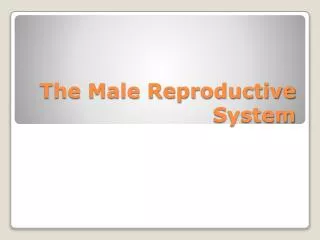
Marieb’s Human Anatomy and Physiology Ninth Edition Marieb w Hoehn. Chapter 27 Male Reproductive System Lecture 18 Part 1: Overview and Primary Sex Organs. Warning!. The video and narration in this presentation concerns the Male Reproductive System

E N D
Marieb’s Human Anatomy and Physiology Ninth Edition Marieb w Hoehn Chapter 27 Male Reproductive System Lecture 18 Part 1: Overview and Primary Sex Organs
Warning! The video and narration in this presentation concerns the Male Reproductive System and uses explicit graphics and terminology dealing with this subject matter. Some material may not be suitable for younger viewers.
Male Reproductive System • There are three main functions of the male reproductive system • Produce and maintain sex cells (sperm) • Transport sperm and supplemental fluids to the female reproductive tract • Secrete male sex hormones • Sex organs can be divided into • Primary sex organs (gonads) = testes (sperm, hormones) • Accessory (secondary) sex organs = internal and external reproductive organs
Male Reproductive System Figure from: Martini, Anatomy & Physiology, Prentice Hall, 2001 Testis → Epididymis → Vas (ductus) deferens → Ejaculatory duct → Urethra
Male Reproductive Organs posterior view Figure from: Hole’s Human A&P, 12th edition, 2010
Descent of Testes Figure from: Hole’s Human A&P, 12th edition, 2010 Descent begins 1-2 months beforebirth under the influence of testosterone Descent is necessary for sperm production Failure of testes to descend = cryptorchidism
Structure of the Testis Figure from: Hole’s Human A&P, 12th edition, 2010 Surrounded by the tunica albuginea – a tough, white, fibrous capsule that encloses each testicle Septa divide each testicle into about 250 lobules Each lobule contains 1-4 highly coiled seminiferous tubules that give rise to sperm Interstitial cells (of Leydig) lie in between seminiferous tubules and secrete male sex hormones Rete Testis
Review of Mitosis and Meiosis Figures from: Martini, Anatomy & Physiology, Prentice Hall, 2001 Mitosis – production of two identical diploid daughter cells Meiosis – production of four genetically varied, haploid gametes
Seminiferous Tubules and Sperm Maturation Figures from: Martini, Anatomy & Physiology, Prentice Hall, 2001 Spermatogonium = stem cell
Spermatogenesis Know the order of events below! Spermatogonium (2n) Primary spermatocyte (2n) Meiosis I Secondary spermatocyte (n) Meiosis II Spermatid (n) Spermiogenesis Spermatozoan (n)
Formation of Sperm Cells Figure from: Hole’s Human A&P, 12th edition, 2010 • Supporting cellsaresustentacular cells • They: • are important in regulating and supporting spermatogenesis • help maintain the blood-testis barrier
Structure of a Sperm Cell Figure from: Hole’s Human A&P, 12th edition, 2010 Only flagellum in human body Mitochondria Enzymes used to penetrate the egg during fertilization
Review • Spermatogenesis • Spermatogonia, 1o spermatocyte, 2o spermatocyte, spermatid, spermatozoan • Is a result of meiotic division • Under the control of FSH • Is guided and regulated by sustentacular cells • Produces 4 haploid gametes (spermatozoa) • Spermatozoa • Head, midpiece, and tail • Acrosomal cap – enzymes use for fertilization • Non-motile when produced – must undergo capacitation