Download
1 / 22
270 likes | 511 Vues
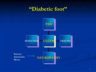
Imaging Assessment of Diabetic Foot Infections. Regina Alivisatos, MD Medical Officer DSPIDPs. Introduction. Patients with osteomyelitis should be identified in order to ensure the most appropriate course of treatment a homogenous clinical trials population

E N D
Imaging Assessment of Diabetic Foot Infections Regina Alivisatos, MD Medical Officer DSPIDPs
Introduction Patients with osteomyelitis should be identified in order to ensure • the most appropriate course of treatment • a homogenous clinical trials population 7 – 14% of enrolled subjects found to have osteomyelitis • excluded from the PP populations • failures in the ITT analysis
WHY? • Decreasing size of the PP populations that may be distributed unequally • Inaccurate assessment of the true efficacy for one or both of the treatment arms • Database size insufficient to draw conclusions about a drugs efficacy in CSST infections or in the diabetic foot subset
Complications • Determination of infection complicated because of superimposed neuropathic osteoarthropathy and peripheral vascular disease • Neuropathic disease can lead to f/x, deformity, bone production, and hyperemia which can mimic infection on MRI and scanning increasing the false positives • Peripheral vascular disease can prevent contrast material or tracer from reaching site of concern and lead to false negatives
Diagnosis - osteomyelitis • Presence of osteomyelitis impacts on failure rate of soft tissue infections • “gold standard” is bone histology and culture through non-infected tissue
Procedures 1) Plain films 2) Radionuclide or Scintigraphic imaging • Triple Phase Bone Scan (TPBS) • Gallium Scan • Indium-111 Leukocyte Scan 3) Magnetic Resonance Imaging (MRI) 4) Probe to Bone
Procedures 1) Plain films 2) Radionuclide or Scintigraphic imaging • Triple Phase Bone Scan (TPBS) • Gallium Scan • Indium-111 Leukocyte Scan 3) Magnetic Resonance Imaging (MRI) 4) Probe to Bone
X-Ray Initial screening tool: • Easily obtained, relatively inexpensive and provides anatomical information • Demineralization, periosteal reaction, bony destruction: (the classic triad) • Appear after 30 – 50% of bone destroyed and can take as much as 2 weeks to appear • Found in other conditions such as fracture or deformity • Sensitivity and specificity approximately 54% and 80%
Procedures 1) Plain films 2) Radionuclide or Scintigraphic imaging • Triple Phase Bone Scan (TPBS) • Gallium Scan • Indium-111 Leukocyte Scan 3) Magnetic Resonance Imaging (MRI) 4) Probe to Bone
Three-phase bone scintigraphy (TPBS) • Highly sensitive since positive as early as 24 hours after onset • Focal hyperperfusion, hyperemia, bony uptake • Can also be seen in fractures, neuropathic joints and longstanding cellulitis decreasing specificity • Fourth phase (24 hour image) enhances specificity • Concurrent TPBS with IN111 scanning optimal
TPBS • Literature review of 20 reports of 1,166 patients (method of confirmation of osteomyelitis diagnosis not specified) • In patients w/o prior bone changes: 94% sensitive and 85% specific for osteomyelitis • In patients with complicating conditions: 95% sensitive, 33% specific. Schauwecker et al; The scintigraphic diagnosis of osteomyelitis. AJR 1992; 158(1):9-18
Gallium Scanning • Must be performed with a TPBS • Diagnostic criteria include • gallium uptake exceeds TPBS scan uptake • gallium and TPBS scan results are discordant • Sensitivity 81% and specificity 69% • Cost of gallium scan AND TPBS may exceed cost of a single more sensitive and specific test such as an Indium scan or an MRI Schauwecker et al. AJR 158; 9 - 18, January 1992
Indium scanning • Best sensitivity, specificity, and cost compromise in patients with and without prior bone abnormalities • Issue of practicality of labeling WBCs and later images • Does not accumulate at sites that are not infected • Compilation of sensitivity and specificity for 142 diabetic subjects from 5 studies showed sensitivity of 88.6% and specificity of 84% Schauwecker et al. AJR 158; 9 - 18, January 1992
Procedures 1) Plain films 2) Radionuclide or Scintigraphic imaging • Triple Phase Bone Scan (TPBS) • Gallium Scan • Indium-111 Leukocyte Scan 3) Magnetic Resonance Imaging (MRI) 4) Probe to Bone
MRI: High-tech, high cost? • Decreased marrow signal intensity on T1-weighted images and increased signal intensity on T2-wighted images with marrow enhancement after injection of contrast • Associated findings of soft tissue mass, cortical destruction, sequestrum formation and sinus tracts with ulceration increase diagnostic certainty • Good anatomical detail • Sensitivity and specificity comparable to that of Indium scan • Review of 129 diabetics showed MRI sensitivity of 86% and specificity of 84% American College of Radiology: Imaging diagnosis of Osteomyelitis in patients with DM/Appropriateness Criteria, 1999
MRI continued • 62 feet in 59 patients with suspected osteomyelitis were prospectively evaluated (27 with DM, 35 w/o) • In DM sensitivity 82%, specificity 80% • In non-DM: sensitivity 89%, specificity 94% • Accuracy increased with contrast-enhanced studies (89%) vs.78% • Cost savings initially because test is more rapid • Competitively priced compared with combination of TPBS and Indium or with gallium • Allows good delineation of surgical field Morrison, WB et al, Radiology; Aug 1995:196:557-64
TPBS with In-111-labeled WBC scintigraphy in the examination of the feet in diabetic patients: Results of Published Reports
Procedures 1) Plain films 2) Radionuclide or Scintigraphic imaging • Triple Phase Bone Scan (TPBS) • Gallium Scan • Indium-111 Leukocyte Scan 3) Magnetic Resonance Imaging (MRI) 4) Probe to Bone
Probe • 75 subjects with 76 ulcers from one center • Osteomyelitis diagnosed in 50 (66%), excluded in 26 • Confirmation based on histologic examination • culture data not analyzed as cultures were taken from base of infected ulcer • if bone biopsy not done, diagnosis was based on radiographic tests or surgeons finding of purulent nonviable bone • Bone probed in 36 of 50 with contiguous osteomyelitis and in 4 of 26 w/o osteomyelitis • Sensitivity 66%, specificity 85%, positive predictive value 89%, negative 56% • Conclusion: Palpation of bone strongly correlated with presence osteo. Probing included in initial assessment of diabetics with infected ulcers. Specialized imaging studies not necessary if positive Grayson et al JAMA 1995 Mar 1;273(9):721-3