Download
1 / 72
720 likes | 727 Vues
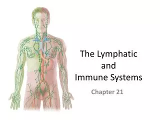
Chapter 20, 21. Lymphatic and Immune Systems. Part I. Tissues and Nonspecific Immunity. Overview. Lymphatic system functions Lymph vessel anatomy Lymphocytes Lymphatic tissues (nodules) Lymphatic organs (nodes, thymus, spleen) Nonspecific defenses. The Lymphatic System.

E N D
Chapter 20, 21. Lymphatic and Immune Systems Part I. Tissues and Nonspecific Immunity
Overview • Lymphatic system functions • Lymph vessel anatomy • Lymphocytes • Lymphatic tissues (nodules) • Lymphatic organs (nodes, thymus, spleen) • Nonspecific defenses
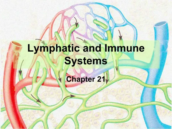
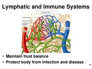
The Lymphatic System • Consists of two semi-independent parts: • A network of lymphatic vessels • Lymphoid tissues and organs scattered throughout the body • Returns interstitial fluid and leaked plasma proteins back to the blood • Lymph = interstitial fluid once it has entered lymphatic vessels • Produces, maintains, and distributes lymphocytes • Protects us against pathogens: microscopic organisms that cause disease: • viruses • bacteria • fungi • parasites • Each attacks in a specific way
Parts of the Lymphatic System • Lymph: • a fluid derived from plasma/interstitial fluid • does not have plasma proteins • Lymphatic vessels(lymphatics): • network that carries lymph from peripheral tissues to the venous system • Lymphoid tissues and lymphoid organs: • found throughout the body • Lymphocytes, phagocytes, and other immune system cells
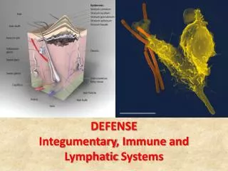
The Immune System • Includes all body cells and tissues involved in production of immunity, not just lymphatic system • What else? Integumentary, digestive, cardiovascular, respiratory, others all contribute to the immune system
Organization of the Lymphatic System Figure 22–1
Lymphocyte Circulation • From blood plasma to interstitial fluid (through capillary exchange) to lymph and back to the venous system
Lymphatic Vessels • One-way system of vessels that carry lymph toward the heart • Lymphatic system begins with smallest vessels called lymphatic capillaries (terminal lymphatics) • Lymphatic caps differ from blood caps in 4 ways: • start as dead end pockets rather than tubes • larger diameters • thinner walls • flat or irregular in section
Lymphatic Capillaries Figure 22–2
Lymphatic Capillaries • Endothelial cells loosely bound together with overlap which acts as one-way valve: • allows fluids, solutes, viruses, and bacteria to enter (very permeable) • prevents return to intercellular space • During inflammation, lymph capillaries can absorb: • Cell debris • Pathogens • Cancer cells • Cells in the lymph nodes cleanse and “examine” this debris • Lacteals = special lymphatic capillaries in small intestine, transport lipids from digestive tract
Lymph Flow • From lymphatic capillaries to larger lymphatic collecting vessels containing larger one-way valves • Valves make them appear beaded • Structure similar to veins • Lymphatic vessels travel with veins
Lymphatic Vessels and Valves Figure 22–3
Superficial and Deep Lymphatic Vessels • Superficial lymphatic vessels are located in: • skin • mucous membranes • serous membranes lining body cavities • Deep lymphatic vessels are larger vessels that accompany deep arteries and veins • Superficial and deep join to form large lymphatic trunks, which empty into 2 major collecting vessels: • thoracic duct • right lymphatic duct
Lymphatic Ducts and the Venous System Figure 22–4
Right lymphatic duct collects lymph from: drains the right upper arm and the right side of the head and thorax; empties into right subclavian vein Thoracic Duct collects lymph from rest of body and empties into left subclavian vein Lymph Return to Venous Blood
Lymph Transport • The lymphatic system lacks a pumping organ • Vessels are low-pressure conduits • Uses the same methods as veins to propel lymph: • Pulsations of nearby arteries • Contractions of smooth muscle in the walls of the lymphatics
Lymphedema • Blockage of lymph drainage from a limb causes severe swelling • Fluid is stagnant, does not get filtered by lymph nodes • Thus, infections are likely and can be very dangerous
Specific Defenses • Lymphocytes: part of the immune response, a specific defense system • Respond to: • environmental pathogens • toxins • abnormal body cells, such as cancers • Detect problems and travel into site of injury or infection • Identify, attack, and develop immunity to a specific pathogen • Immunity: the ability to resist infection and disease
Lymphocyte Production • Lymphocytes are produced in: • lymphoid tissues (e.g., tonsils) • lymphoid organs (e.g., spleen, thymus) • red bone marrow • Make up 20–30% of circulating leukocytes • Majority are stored in lymphoid organs, not circulating (remember that only about 1% of your WBCs are in the blood)
Classes of Circulating Lymphocytes • T cells: thymus-dependent • Make up 80% of circulating lymphocytes • B cells: bone–marrow derived • Make up 10–15% of circulating lymphocytes • NK cells: natural killer cells • Make up 5–10% of circulating lymphocytes
Types of T Cells • Cytotoxic T cells • Attack cells infected by viruses • Produce cell-mediated immunity • Helper T cells • Stimulate function of T cells and B cells • Suppressor T cells • Inhibit function of T cells and B cells • Inflammatory T cells • The last two are called regulatory T Cells because they control the sensitivity of immune response
B Cells • Differentiate into plasma cells, which produce and secrete antibodies(immunoglobin proteins) • Antibodies bind to their specific target antigen and initiate antibody-mediated immunity
Antigens • Antigen – anything the body perceives as foreign • Bacteria and their toxins; viruses • Mismatched RBCs or cancer cells
Natural Killer (NK) Cells • Also called large granular lymphocytes • Responsible for immunological surveillance • Attack: • foreign cells • virus-infected cells • cancer cells
Lymphocyte Distribution • Tissues maintain different T cell and B cell populations • T Cells: high in blood, thymus, marrow, spleen, others • B Cells: high in nodes, spleen, others • Lymphocytes wander through tissues, migrating throughout the body to defend peripheral tissues (T cells move faster than B) • They can enter blood vessels or lymphatics for transport • Have long life span (4 years+, up to 30!) • Retain their ability to divide, which is essential to immune system function
Lymphopoiesis • Lymphocyte production involves: • bone marrow • thymus • peripheral lymphoid tissues Figure 22–5
Hemocytoblasts • In bone marrow, divide into 2 types of lymphoid stem cells • Group 1: • remain in bone marrow • produce B cells and natural killer cells • B cells differentiate with exposure to cytokine (immune system hormone) produced in the bone marrow called interleukin 7 (IL-7) • Group 2: • migrate to thymus • produce T cells in environment isolated by blood-thymus barrier • Differentiate in response to thymic hormones (thyomsins)
Lymphoid Functions • Lymphoid tissues and lymph nodes: • Distributed throughout body to monitor peripheral infections respond before infections reach vital organs of trunk • Lymph nodules in mucosa • Lymph nodes monitor plasma/interstitial fluid
Lymphoid Tissue • Diffuse lymphatic tissue – scattered reticular tissue elements in every body organ • Larger collections appear in the lamina propria of mucous membranes and lymphoid organs • Lymphoid follicles (nodules) – solid, spherical bodies consisting of tightly packed reticular elements and cells • Germinal center composed of dendritic and B cells • Found in isolationand as part of larger lymphoid organs
Lymphoid Nodules • Areolar tissue with densely packed lymphocytes • Germinal center contains dividing B cells Figure 22–6
Distribution of Lymphoid Nodules • Respiratory tract (tonsils) • Along digestive tract (MALT = Mucosa Associated Lymphoid Tissue e.g. Peyer’s patches, appendix) • Urinary tract • Found within some lymphoid organs (Lymph nodes, spleen)
Lymphoid Organs • Are separated from surrounding tissues by a fibrous connective-tissue capsule = encapsulated. • Include: • Lymph nodes • Thymus • Spleen
Lymph Nodes Diameter = 1-25 mm Figure 22–7
Lymph Node • Principal lymphoid organs of the body • Embedded in connective tissue and clustered along lymphatic vessels • Aggregations of these nodes occur near the body surface in inguinal, axillary, and cervical regions of the body (Lymph Glands) • Functions • Act as filters, purifying lymph before returning it to venous circulation, removes debris, pathogens, 99% of antigens • monitor for antigens and mount an attack against them (activate the immune system)
Lymph Node - Structure • Surrounded by fibrous capsule • Trabeculae: fibrous partitions made of collagen fibers that extend from capsule into interior of lymph node • Hilus: shallow indentation where blood vessels and nerves reach the lymph node • Afferent Lymphatic Vessels: carry lymph from peripheral tissues to lymph node • Efferent Lymphatic Vessels: leave lymph node at hilus, carry lymph to venous circulation
Lymph Flow • Flows from afferent lymphatics through lymph node in a network of sinuses: • Enters subcapsular sinus: • contains macrophages and dendritic cells • Through outer cortex: • contains B cells within germinal centers • Through deep cortex: • dominated by T cells • Through the core (medulla): • contains B cells and plasma cells • organized into medullary cords • Into hilus and efferent lymphatics (less of theses than afferent)
Antigen Presentation • First step in immune response • Extracted antigens are “presented” to lymphocytes by macrophages, dendritic cells (in lymph nodes, these are in the subcapsular area)
Lymphoid Organs • The spleen, thymus gland, and tonsils • Peyer’s patches and bits of lymphatic tissue scattered in connective tissue • All are composed of reticular connective tissue • All help protect the body • Only lymph nodes filter lymph Figure 20.5
The Thymus Figure 22–8
The Thymus • Secretes hormones (thymosin and thymopoietin) that cause T lymphocytes to mature • Located in mediastinum • Deteriorates after puberty • Divided into 2 thymic lobes • Septa divide lobes into smaller lobules • Each lobule contains: • a dense outer cortex of dividing T cells • a pale central medulla
Internal Anatomy of the Thymus • Thymic lobes contain an outer cortex and inner medulla • Cortex contains densely packed lymphocytes and scattered macrophages. Cortex cells: • Surround lymphocytes • Maintain blood-thymus barrier • Secrete thymic hormones (thymosins) that stimulate stem cell divisions and T cell differentiation • Medulla contains fewer lymphocytes and thymic (Hassall’s) corpuscles. Medulla cells: • Form concentric layers (Hassall’s corpuscles) ?? • The medulla has no blood–thymus barrier: here T cells can enter or leave bloodstream
Thymus • The thymus differs from other lymphoid organs in important ways • It functions strictly in T lymphocyte maturation • It does not directly fight antigens • The stroma of the thymus consists of star-shaped epithelial cells (not reticular fibers) • These thymocytes secrete the hormones that stimulate lymphocytes to become immunocompetent • T cells: • migrate into medulla • divide in the cortex • leave thymus by medullary blood vessels
The Spleen Figure 22–9
Spleen • Largest single collection of lymphoid tissue in the body, located on left side of abdomen beneath the diaphragm • Filters the blood like lymph nodes filter the lymph • Phagocytes and other lymphocytes in the spleen identify and attack damaged and infected cells in circulating blood • Functions: • Site of lymphocyte proliferation • Immune surveillance and response • Cleanses the blood
Functions of the Spleen • Removal of abnormal blood cells and other blood components by phagocytosis • Storage of iron and other RBC products for later use or elimination • Initiation of immune responses by B cells and T cells in response to antigens in circulating blood • Site of lymphocyte proliferation • Stores blood platelets
Structure of the Spleen • Splenic veins, arteries, and lymphatic vessels all communicate with spleen at hilus • Inside fibrous capsule (with trabeculae): • red pulp: • contains many worn out red blood cells, macrophages • Area of RBC disposal • white pulp: • contains many WBCs • resembles lymphoid nodules
Splenic Circulation • Splenic artery branches into Trabecular Arteries • Branch and radiate toward capsule • Finer branches surrounded by white pulp • Capillaries discharge red blood cells into red pulp • Contains elements of circulating blood plus fixed and free macrophages • Blood passes through a network of reticular fibers • Then enters large sinusoids (lined by macrophages) which empty into trabecular veins
Structure of the Spleen Figure 20.6a, b
Immunity: Two Intrinsic Defense Systems • Innate (nonspecific) system responds quickly and consists of: • First line of defense – skin and mucosae prevent entry of microorganisms • Second line of defense – antimicrobial proteins, phagocytes, and other cells • Inhibit spread of invaders throughout the body • Inflammation is its most important mechanism • Adaptive (specific) defense system • Third line of defense – mounts attack against particular foreign substances • Takes longer to react than the innate system • Works in conjunction with the innate system
Innate and Adaptive Defenses Figure 21.1