Download
1 / 32
330 likes | 530 Vues
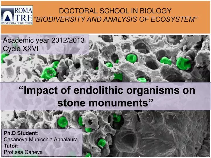
DOCTORAL SCHOOL IN BIOLOGY “BIODIVERSITY AND ANALYSIS OF ECOSYSTEM”. Academic year 2012/2013 Cycle XXVI. “Impact of endolithic organisms on stone monuments”. Ph.D Student : Casanova Municchia Annalaura Tutor: Prof.ssa Caneva. Biodeterioration/ endolithic microorganisms. Introduction.

E N D
DOCTORAL SCHOOL IN BIOLOGY “BIODIVERSITY AND ANALYSIS OF ECOSYSTEM” Academicyear2012/2013 Cycle XXVI “Impact of endolithic organisms on stone monuments” Ph.DStudent: Casanova MunicchiaAnnalauraTutor: Prof.ssa Caneva
Biodeterioration/ endolithic microorganisms Introduction Endolithic growth form can be present in different microorganisms groups: cyanobacteria, green and red algae, fungi, and lichens. Their grow inside the stone in order to protect themselves from adverse conditions (high solar radiation, adverse temperature and desiccation conditions) Can penetrate some millimeters or centimeters inside the rock
Biodeterioration/ endolithic microorganisms Introduction The stone monuments, as well as rocks, can be colonized by endolithic microorganisms showing biodeterioration phenomenon. Often it is not recognized and is confused with abiotic cause Understimated question despite is the most dangerous biological deterioration processes affecting stone monuments. Morphology of pitting from the Trajan Column. Caneva et al., 1994
Aims GeneralAim: A major contribution about the knowledge of biodeterioration process due to endolithson the stone monuments by discovering new approaches and new techniques In Detail : Detection of endolithic traces on stone monuments in Temperate and Mediterranean bioclimates Microorganismsadoptorganic or inorganicsurvivalstrategies under high stress conditions, leavingbiological or geological traces on rock Study of the spatiality of endolithicmicroorganisms in relation with the stone
Samples Materials and Methods Natural outcrops Stone monuments (Case studies) 7 samples – Marble rock from Carrara area. 1 sample- Black schistHebrew’s cemetery tombstone in Venice. 3 samples- Dolomitic limestone cliff of the Amalfi Coast. 11 samples- Carbonate limestone Church in Martvilli (Georgia)
Optical Microscope/ SEM-EDS Materials and methods Experimental protocol Observation of polished cross-section Spread and the depth of the colonization Observation of polished sections after staining with Periodic Acid Schiff (PAS)
Optical Microscope/ SEM-EDS Materials and methods Experimental protocol Observation in light trasmitted Observation of thin-section Morphology of microorganisms and bioalteration induce on stone SEM observation of cross-section after acid attack and fractured sample
RamanSpectroscopy Materials and methods Raman spectroscopy is an analytical technique that provides molecular structural information, based on inelastic scattering of monochromatic light. to identify traces of the organic and inorganic compounds left by endolithic microorganisms
Confocal Laser Scanning Microscope Materials and methods Provide series of thin optical sections of the sample, at different intervals along the Z axis 3-D image of fluorescent organisms or stained with fluorescent labels to determine the occurrence, the spatial organization and the volume of endolithic microorganisms
ImageAnalysis Materials and methods Display, analyze, process the image obtained by CLSM. ImageJ® Java-based image processing program Quantify the bio-volume occupied inside the stone of each stack , develop profiles and isosurfaces in 3-Dimension. ImageJ Plugin 3D viewer and voxels counter Imaris software ,Trial version, (Bitplane).
Papers • V. Lombardozzi, T. Castrignanò, M. D’Antonio, A. Casanova Municchia, G. Caneva, An interactive database foranecologicalanalysisofstonebioppitting, Int. Biodeterioration & Biodegradation,2012, 73, 8. • 2. G. Caneva, V. Lombardozzi, S. Ceschin, A. Casanova Municchia, O. Salvadori, Unusual differential erosion related to the presence of endolithic microorganisms ( Martvili, Georgia), Journal of Cultural Heritage, 2013,in Press • 3. A. Casanova Municchia, G. Caneva, M. A. Ricci, A. Sodo, Identification of endolithic traces on stone monuments, in review to the Journal of Raman Spectroscopy • 4. A. Casanova Municchia, Z. Percario, G. Caneva, • Detection of endolithic spatial distribution in marble stone using Confocal laser scanning microscopy,submitted to the Journal of Microscopy • .
An interactive database for an ecological analysis of stone biopitting Papers1/4 Interactive online database developed in order to : -Synthesize the available information on the stone-pitting phenomena -Identify the trends of stone-pitting phenomena, the most favorable environmental conditions, the most affected kind of stone, the most common biodeteriogens. 24 are the papers used to building up the database; 83 the total number of sites ; 249 the total number samples. Most of the sampling sites are in the Mediterranean Basin
Results/Conclusions Papers1/4 Marble is often described as the most affected material On carbonate and marble rocks is found the most evidence of appearance of cyanobacteria Cyanobacteria are the dominant group associated with pitting Biopitting is mainly described in vertical and subvertical surfaces, showing a preference for southern exposures
Unusual differential erosion related to the presence of endolithic microorganisms (Martvili, Georgia) Papers2/4 A differential erosion phenomenon was observed on the walls of the Church of the Virgin in Martvilli. Characterized by the circular imprints left in the stone (from 1 cm to 3 cm in diameter) Detail of the stone surface affected by biodeterioration phenomena on South-facing side of the Church. AIMS - Provides an interpretation of the differential erosion phenomenon - Identify the ecological conditions which favor the phenomenon -Impact on the stone conservation
Results Papers2/4 The stone is a fine-grained limestone. Traces of fossils with relatively high values of porosity (23.28%). The southern facade is the most intensely affected by the differential erosion phenomenon Cyanobacteria are the most common microorganisms occuring Through the analysis of images it was possible to estimate a considerable colonized area The microorganisms appears to from an average depth of 200 μm Black meristematic fungi are on and below the surface of the sample
Conclusion Papers2/4 This unusual differential erosion phenomenon is related to : -The physical petrographic features of the rock, (heterogeneity and discontinuity) giving rise the deterioration phenomenon in specific preferential areas. - Intense xeric conditions that permit the establishment of endolithic microorganisms (Southern facade ) -Biodeterioration due to the cyanobacteria and meristematic fungi endolithic activity
Identification of endolithic traces on stone monuments Papers3/4 Raman spectroscopic analysis applied to four endolithic samples from Temperate and Mediterranean bioclimate regions Oral Presentation: 7th International Conference on the application of Raman spectroscopy in Art and Archaeology,Ljubljana 2-6 September 2013 to identified the traces of organic and inorganic compounds present in the stone monuments High stress conditions inducing the microorganisms to adopt survival strategies protection from desiccation and high solar radiation Scytonemin UV-protection Calcium oxalates Aridity tolerance
Materials and Methods Papers3/4 Four samples colonized by endoliths -Marble rock showing colonization of cyanobacteria and endolithic fungi from quarries in Carrara Area -Black schist from Hebrew’s cemetery tombstone in Venice. Endolithic lichens with perithecia completely sunken in the rock; - Dolomitic limestone showing colonization of endolithic cyanobacteria from cliff of the Amalfi Coast -Fine-grained limestone from the Church of the Virgin in Martvili in Western Georgia. The back side shows orange biological traces
Materials and Methods Papers3/4 All measurements have been performed with a Renishaw In‑Via Reflex Raman microscope 785 nm near-infrared and the green laser line at 514 nm; objectives 50X; Raman spectra have been recorded on the surface and in the inner part of the samples, to identify a possible difference of the bio- and geotraces detected at different depth
Results/ Sample 1 Papers3/4 Calcite Scytonemin Marble rock Inner side Scytonemin is synthesized by cyanobacteria as extracellular sheath pigment, against UV radiation Anthraquinonecoumpounds against intense solar radiation Chlorophyll Surface
Results/ Sample 2 Papers3/4 Fe Black schist from Hebrew’s cemetery • Goethite • α-FeO(OH) -Lepidocrocite δ-FeO(OH) Carbon-based substance Dolomite Calcite
Results Papers3/4 Black schist from Hebrew’s cemetery Anthraquinone coumpounds against intense radiation
Results/Sample 3 and Sample 4 Papers3/4 Raman spectra recorded using the 785 nm laser line, show only the substrate 514 nm laser excitation to identify the organic traces typical spectrum of a carotenoid with bands centered at 1522 and 1154 cm−1. Dolomitic limestone Carotenoid is an accessory pigment, usually produced under stress conditions within antioxidant strategy limestone from the Church of the Virgin in Martvili
Conclusions All samples show traces of compounds known to be effective against UV-radiation damage -Sample1-Marble from Carrara area. Scytoneminappears in all spectra and at different depths of the sample. Anthraquinonecoumpounds -Sample2-Hebrew’s cemetery tombstone in Venice. Iron oxide hydroxides due to a bioalteration by endolithic. Anthraquinonecoumpounds - Sample3-4 Dolomitic limestone and sample from Church in Martvili Carotenoid compounds (antioxidant) useful against the high UV-radiation
Detection of endolithic spatial distribution in marble stone using Confocal laser scanning microscopy Papers4/4 Aims Detection of the endolithic spatial distribution and quantify the bio-volume occupied using the confocal laser scanning microscopy (CLSM) with a double- staining. Understand the real impact of on the stone conservation Compare the results with those acquired from microscopy techniques (SEM and light microscope)
Materials and Methods Papers4/4 Rock flakes from the Marble rock samples stained with : The nucleic acid stain, propidium iodide ( after permeabilization of the cell membranes) DNA structures excitation 543 nm emission 633 nm ( RED CHANNEL ) The glycoconjugates stain lectinConcanavalin-A Alexa Fluor 488 Extrapolymeric substances (EPS) excitation 543 nm emission 633 nm ( GREEN CHANNEL) The CSLM images were collected in a set of optical cross- sectional image in the x-y plane obtained at different intervals along the z-axis. ( Total Z path 160 μm)
Results Papers4/4 Isosurface presentations show the 3-D arrangement 760 μm Microorganisms stained with red fluorescent (PI) Extracellular matrix (EPS) stained with green fluorescence (ConA- Alexa Fluor 488) Overview in y-x directions shoving the penetrations Overview in x-z directions shoving the penetrations in the sample thickness Overview in x-z direction of the cyanobacteria distribution
Results Papers4/4 Volume distribution; ConA- Alexa Fluor 488 stain Volume distribution; propidium iodide stain Total Volume 2 % Total Volume 2.5 % Volumes were calculated from each of six image stacks corresponding to the various depths
Results/ Comparison PAS and SEM Papers4/4 Entire cross-section partially decalcified by hydrochloric acid solution 3 mm Cyanobacteria immersed in the substratum Total Area 6 % Calcite grains perforated by hyphae Fungal hyphae embedded in extracellular matrix (white arrow) Cross-section after PAS staining After application of a threshold classification, the substratum (black area) and endolithic colonization (white areas).
Conclusion Papers4/4 The marble rock sample is colonized by cyanobacteria completely immersed in the stone. Below the cyanobacteria a dense network of fungal hyphae is present CLSM with the double staining distinguish and quantify the contribution of the extracellular matrixfrom that of DNA structure The volume occupies by endolithiccyanobacteria and fungi is about 2.5% and the EPS Volume is about 2 % Microscope CSLM results provided a good information about the 3-D spatiality of the endolithic microorganisms, the real volume occupied, the distribution between the grains and the penetration into the calcite grains. This is the first study of the biodeterioration phenomenon due to endolithic microorganism aimed at stone monuments by the use of CLSM microscope with double staining
GeneralConclusion A increase of the information about the real effect on the stone monuments andof the potential damage by endolithic attack New approaches and new techniques have been applied A prove of the potentiality of the Raman Spectroscopy, which is here applied for the first time on stone monuments, in the identification of traces of biological compounds from endolithic microorganisms A useful contribution for a clear identification of the presence of endolithic microorganisms on stone monuments A new approach, with the employment of 3-D technologies, in the evaluation the real impact of endolithic microorganisms on the stone monuments
I wishtothanks: Di Giulio A. ; Ricci M.A.; Salvadori O; Sodo A.; PercarioZ.; and the LIME staff for the technicalsupport. I thankall the doctoral school and all the Ph.D students A big thank to my tutor Prof.ssa G. Caneva for her help and for all the experiences of this three years and I wantover-please the laboratoryfor the support and the best moments (Alma, Roberto; Francoise, Flavia, Valentina; Giulia, Simona, Wawan). Thanksforyourattention…