Download
1 / 12
120 likes | 311 Vues
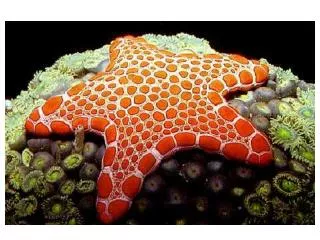
Dissection of a Sea Star. Adult Sea Stars have penta-radial symmetry; therefore, each ray or arm contains the same organ structures as the other arms. The structures in the central disc are not in multiples of five. Photos by Dr. J. Whaley Flagler Palm Coast High School 2013.

E N D
Dissection of a Sea Star Adult Sea Stars have penta-radial symmetry; therefore, each ray or arm contains the same organ structures as the other arms. The structures in the central disc are not in multiples of five. Photos by Dr. J. Whaley Flagler Palm Coast High School 2013
External Anatomy – Aboral Surface (Dorsal Surface) Central Disc- at the center the the disc is the anus Arm or Ray Madreporite (or seive plate)
External Anatomy – Aboral Surface Madreporite Located on the central disc at the junction between two of the arms. Functions to draw water into the water vascular system of the sea star. The larger, longer projections are the spines, which aid in protection. The smaller projections surrounding the spines are the skin gills which function to take in oxygen and the pedicellariae (small pincher-like structures that help keep the skin clear of debris.) Spine Pedicellariae Skin Gills
External Anatomy – Oral Surface (Ventral Surface) Tube Feet Mouth Ambulacral Groove
External Anatomy – Oral Surface Note the rows of tube feet in the ambulacral groove. The tube feet function in locomotion, feeding, respiration and excretion.
Internal Anatomy After careful removal of the dorsal surface (aboral surface) of skin from one of the arms of a sea star, you may notice the network of structures located directly under the skin. These are the ossicles, that help make up the endoskeleton. They are closely associated with the spines. Ossicles
Internal Anatomy Located under the skin and endoskeleton in the arms of a sea star are the digestive glands. They produce digestive enzymes which will help breakdown food substances. These glands appear to take up most of the arm of the sea star. Note the vessels which connect the digestive glands to the stomach region. Vessels leading to the stomach Digestive Glands Stomach To see the stomach, the skin over the central disc has to be carefully removed. When the skin is removed, the connection to the anus is also cut. The dorsal portion of this region is the pyloric stomach. It is hard to distinguish from the cardiac stomach which lies below it.
Internal Anatomy When the stomach is removed, it should be examined for the presence of any food substances. In some specimens, the cardiac stomach may be partially everted through the mouth. Remember, the sea star will evert its stomach when eating. Stomach (as seen from oral surface)
Internal Anatomy When the digestive glands are moved to the side, the paired gonads are visable . They are located in the arms, close to the central disc. You cannot visually distinguish between the ovaries and testes. Digestive Glands Gonads, paired
Internal Anatomy – Water Vascular System Water enters the Madreporite on the aboral surface. From there, water travesl down the Stone Canal to the Ring Canal. Water then enters each arm through the Radial Canals. Lateral Canals (not visible) connect the Radial Canals to the Ampullae. The Ampullae are bulb- like structures which connect to the tops of the tube feet. Madreporite Stone Canal - cut Radial Canal
Internal Anatomy – Water Vascular System Ring Canal Radial Canals
Internal Anatomy – Water Vascular System Ossicles Ampullae Radial Canal