Download
1 / 1
10 likes | 140 Vues
Biophysical methods analysis on chromatin Fast neutrons protection of chromatin by Cs+ and Al3+ ions, analyzed by static spectrofluorimetry and time resolved spectroscopy Radulescu I, Preoteasa V, Radu L, Serbanescu A, Constantinescu.

E N D
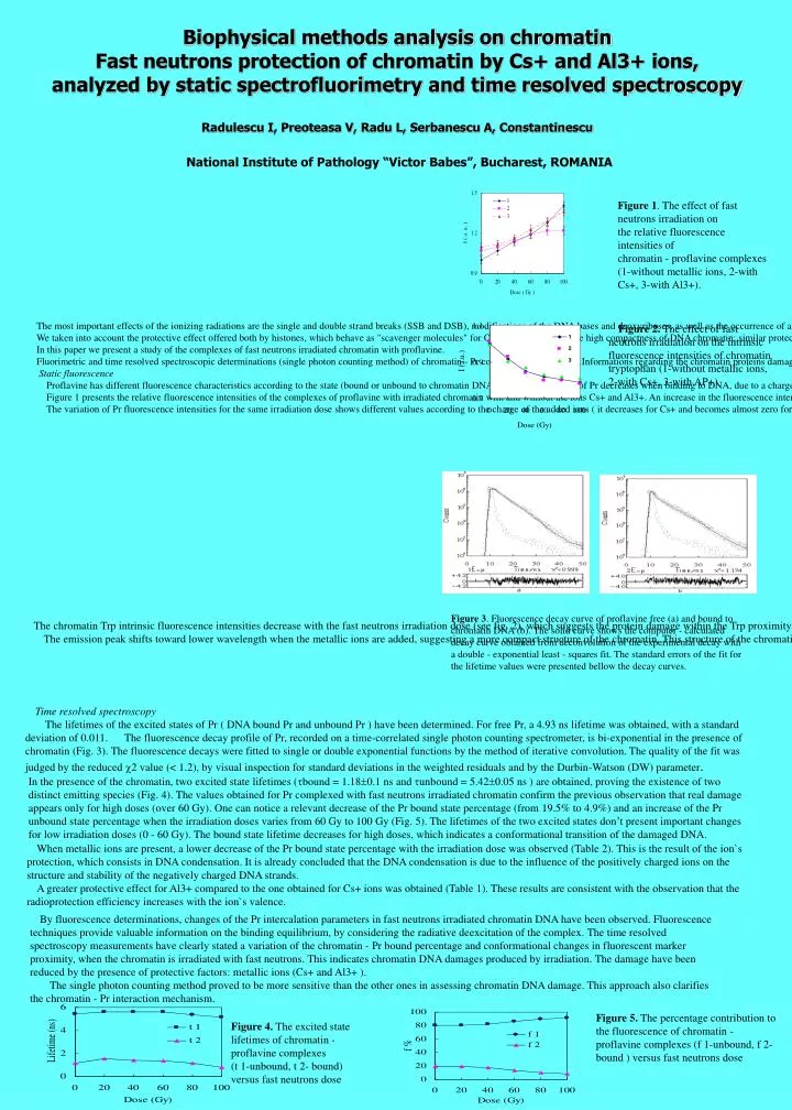
Biophysical methods analysis on chromatinFast neutrons protection of chromatin by Cs+ and Al3+ ions, analyzed by static spectrofluorimetry and time resolved spectroscopyRadulescu I, Preoteasa V, Radu L, Serbanescu A, Constantinescu National Institute of Pathology “Victor Babes”, Bucharest, ROMANIA The most important effects of the ionizing radiations are the single and double strand breaks (SSB and DSB), modifications of the DNA bases and deoxyriboses, as well as the occurrence of alkali and heat labile sites (revealed as strand breaks after alkaline or thermic treatment of irradiated DNA). The ionizing particles can have either direct effects on the DNA constituents or indirect effects, mediated by the OH·_ radicals, appeared as products of the water radiolysis. The occurrence of SSB and DSB in the chromatin DNA strands is supposed to hinder the DNA-dye complex formation. Usually, the dyes present different fluorescence parameters in the two possible states, so one can correlate the lifetime or the quantum yield with the extent of the damage. We taken into account the protective effect offered both by histones, which behave as "scavenger molecules" for OH·_ radicals and by the high compactness of DNA chromatin; similar protective effects might be the result of the metallic ion addition, which triggers some conformational transitions of the chromatin DNA toward a highly compacted structure. In this paper we present a study of the complexes of fast neutrons irradiated chromatin with proflavine. Fluorimetric and time resolved spectroscopic determinations (single photon counting method) of chromatin - Pr complexes were realized.Informations regarding the chromatin proteins damage were obtained by monitoring the fluorescence of Trp. Static fluorescence Proflavine has different fluorescence characteristics according to the state (bound or unbound to chromatin DNA). The quantum yield of Pr decreases when binding to DNA, due to a charge transfer from adenine to the dye. The result is a lower quantum yield of DNA-Pr complex, compared to that of free Pr. Figure 1 presents the relative fluorescence intensities of the complexes of proflavine with irradiated chromatin with and without the ions Cs+ and Al3+. An increase in the fluorescence intensity, which reflects a lower bound Pr ratio consecutive to the chromatin DNA irradiation is observed. This behaviour is explained by the less binding of Pr to chromatin damaged DNA. A negative relationship between bound Pr ratio and the irradiation dose is observed. An important decrease of the ratio is produced only for high irradiation doses, due to the protective role of the histones and of the high compactness of the DNA within the chromatin. The variation of Pr fluorescence intensities for the same irradiation dose shows different values according to the charge of the added ions ( it decreases for Cs+ and becomes almost zero for Al3+ ) (Fig. 1). The protective effect of metallic ions is observed. Figure 1. The effect of fast neutrons irradiation on the relative fluorescence intensities of chromatin - proflavine complexes (1-without metallic ions, 2-with Cs+, 3-with Al3+). Figure 2. The effect of fast neutrons irradiation on the intrinsic fluorescence intensities of chromatin tryptophan (1-without metallic ions, 2-with Cs+, 3-with AP+) The chromatin Trp intrinsic fluorescence intensities decrease with the fast neutrons irradiation dose (see fig. 2), which suggests the protein damage within the Trp proximity and also the appearance of a non radiative deexcitation process. Also, a redshift (about 9 nm) of the emission peak occurs; one can infer a looser structure of the protein and perhaps of the whole chromatin after irradiation. The emission peak shifts toward lower wavelength when the metallic ions are added, suggesting a more compact structure of the chromatin. This structure of the chromatin induced by the metallic ions denotes also a less uniform distribution of the DNA within the chromatin (i.e. high DNA concentration clusters appears). Little variations of Trp relative fluorescence intensities of irradiated chromatin in metallic ions presence are produced (Fig. 2). Figure 3. Fluorescence decay curve of proflavine free (a) and bound to chromatin DNA (b). The solid curve shows the computer - calculated decay curve obtained from deconvolution of the experimental decay with a double - exponential least - squares fit. The standard errors of the fit for the lifetime values were presented bellow the decay curves. Time resolved spectroscopy The lifetimes of the excited states of Pr ( DNA bound Pr and unbound Pr ) have been determined. For free Pr, a 4.93 ns lifetime was obtained, with a standard deviation of 0.011. The fluorescence decay profile of Pr, recorded on a time-correlated single photon counting spectrometer, is bi-exponential in the presence of chromatin (Fig. 3). The fluorescence decays were fitted to single or double exponential functions by the method of iterative convolution. The quality of the fit was judged by the reduced 2 value (< 1.2), by visual inspection for standard deviations in the weighted residuals and by the Durbin-Watson (DW) parameter. In the presence of the chromatin, two excited state lifetimes (bound = 1.180.1 ns and unbound = 5.420.05 ns ) are obtained, proving the existence of two distinct emitting species (Fig. 4). The values obtained for Pr complexed with fast neutrons irradiated chromatin confirm the previous observation that real damage appears only for high doses (over 60 Gy). One can notice a relevant decrease of the Pr bound state percentage (from 19.5% to 4.9%) and an increase of the Pr unbound state percentage when the irradiation doses varies from 60 Gy to 100 Gy (Fig. 5). The lifetimes of the two excited states don’t present important changes for low irradiation doses (0 - 60 Gy). The bound state lifetime decreases for high doses, which indicates a conformational transition of the damaged DNA. When metallic ions are present, a lower decrease of the Pr bound state percentage with the irradiation dose was observed (Table 2). This is the result of the ion`s protection, which consists in DNA condensation. It is already concluded that the DNA condensation is due to the influence of the positively charged ions on the structure and stability of the negatively charged DNA strands. A greater protective effect for Al3+ compared to the one obtained for Cs+ ions was obtained (Table 1). These results are consistent with the observation that the radioprotection efficiency increases with the ion`s valence. By fluorescence determinations, changes of the Pr intercalation parameters in fast neutrons irradiated chromatin DNA have been observed. Fluorescence techniques provide valuable information on the binding equilibrium, by considering the radiative deexcitation of the complex. The time resolved spectroscopy measurements have clearly stated a variation of the chromatin - Pr bound percentage and conformational changes in fluorescent marker proximity, when the chromatin is irradiated with fast neutrons. This indicates chromatin DNA damages produced by irradiation. The damage have been reduced by the presence of protective factors: metallic ions (Cs+ and Al3+ ). The single photon counting method proved to be more sensitive than the other ones in assessing chromatin DNA damage. This approach also clarifies the chromatin - Pr interaction mechanism. Figure 5. The percentage contribution to the fluorescence of chromatin - proflavine complexes (f 1-unbound, f 2-bound ) versus fast neutrons dose Figure 4. The excited state lifetimes of chromatin - proflavine complexes (t 1-unbound, t 2- bound) versus fast neutrons dose