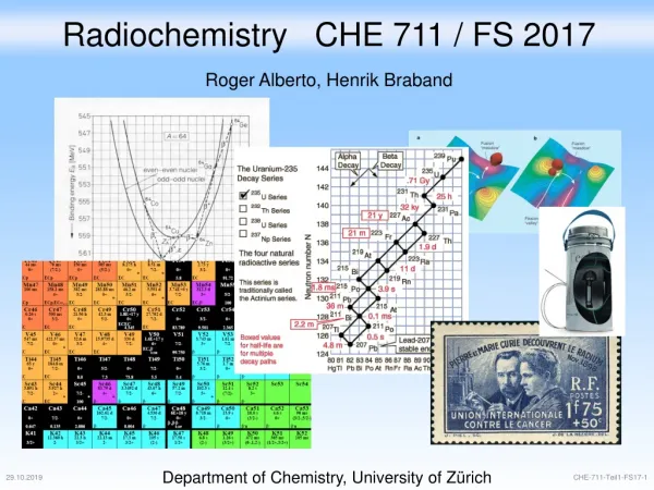
Download
1 / 33
330 likes | 348 Vues
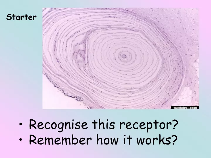
Starter. Recognise this receptor? Remember how it works?. The Pacinian corpuscle is an example of a primary receptor (a single neurone with ONE end detecting stimulus) Pacinian corpuscles function individually. Pacinian corpuscle.

E N D
Starter • Recognise this receptor? • Remember how it works?
The Pacinian corpuscle is an example of a primary receptor (a single neurone with ONE end detecting stimulus) • Pacinian corpuscles function individually.
Pacinian corpuscle Respond to change in mechanical pressure. As with all sensory receptors, a Pacinian corpuscle: • Is specific to a single type of stimulus (e.g. pressure) • Produces a generator potential by acting as a transducer – converts stimuli into a response that can be understood by the body, namely nerve impulses.
Pacinian corpuscle The stimulus is always some form of energy, e.g. heat, light, sound or mechanical energy. The nerve impulse is a form of energy All receptors convert the energy of the stimulus into a nerve impulse known as a generator potential.
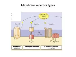
Stretch-mediated sodium channels (Permeability change when shape changes) Resting Potential: Positive outside – negative inside Pressure: Distorts & opens Na+ channels Action potential: Inflow of Na+ depolarises membrane
Pacinian corpuscle In it’s normal (resting) state, the stretch-mediated sodium channels of the membrane around the neurone of a pacinian corpuscle are too narrow to allow sodium ions to pass along them. In this state, the neurone of the pacinian corpuscle has a resting state. When pressure is applied to the corpuscle, it changes shape and its neurone becomes stretched
Pacinian corpuscle This stretching widens the sodium channels in the membrane and sodium ions diffuse into the neurone. The influx of sodium ions changes the potential of the membrane (depolarised), thereby producing a generator potential The generator potential in turn creates an action potential (nerve impulse) that passes along the neurone and then, via other neurones to the CNS.
Receptors working together in the eye Receptors respond to only one type of stimulus. It also only responds to a certain intensity of stimulus. This means the body must have a range of receptors, each responding to a different intensity of stimulus.
Retinal Transduction • The eye is a transducer • What does this mean?
Transduction • The eye is a transducer • What does this mean? • Converts light into a pattern of nerve impulses • Transduction takes place in the retina by a layer of photosensitive cells at the back of the eye • Rods and cones attached to nerves
Photoreceptors These are found in the retina. There are two types Rods and Cones and they are arranged as shown: Outer ! Inner Light Pigmented Layer Ganglion Cells Rod To Optic Nerve Bipolar Neurones Cone
Rod cells Cannot distinguish different wavelengths of light and therefore produce images only in black and white. Rod cells are more numerous than cones.
Rod cells Many rod cells share a single sensory neurone. Rod cells can therefore respond to light of very low intensity. This is because a certain threshold value has to be exceeded before a generator potential is created in the bipolar cells to which they are attached.
Rod cells A number of rod cells are attached to a single bipolar cell (= retinal convergence), there is a much greater chance that the threshold value will be exceeded than if only a single rod cell were attached to each bipolar cell. As a result, rod cells allow us to see in low light intensity (i.e. at night), although only in black and white.
Changes in the electrical potential of a receptor when stimulated by three separate stimuli. Only the third stimulus produces a generator potential high enough to trigger a nerve impulse.
A rod cell LIGHT opsin Rhodopsin (pigment in rod cells broken down) Signal from Bipolar cell
A rod cell As many rod cells are joined to the same bipolar cells, only a single impulse will be stimulated. This means that they cannot distinguish between the separate sources of light that stimulated them. 2 dots close together will appear as a single blob. Rod cells therefore have low visual acuity.
Check! • Discuss with the person sat next to you, the difference between visual acuity and nerve convergence.
How do rod cells produce impulses? Rod cells allow vision in dim light due to the presence of a pigment called rhodopsin, which is found in membrane-bound vesicles. vesicles containing rhodopsin When rhodopsinabsorbs light it splits into its constituent parts, opsin and retinal. This is called bleaching. Low intensity light is sufficient to cause this breakdown. The presence of opsin causes a change in the permeability of the rod cell to sodium, which initiates a generator potential. Rhodopsin can reform in the absence of further light stimulation.
Cone cells Cone cells are of three different types, each responding to a different wavelength of light. Depending on the proportion of each type that is stimulated, we can perceive images in full colour. Each cone cell usually has its own bipolar cell connected to a sensory neurone. This means that often the generator potential is not exceeded. As a result, cone cells only respond to high light intensity and not to low light intensity.
Cone cells Cone cells contain a different pigment to rod cells (iodopsin). This requires a higher light intensity to be broken down and create a generator potential. As cone cells are attached to their own bipolar cell, if 2 adjacent cells are stimulated, the brain receives 2 separate impulses. Cone cells give very accurate vision, they have good visual acuity.
How do cone cells produce impulses? Cone cells aresensitive to high light intensities due to the presence of the pigment iodopsin. vesicles containing iodopsin In bright light, iodopsin is broken down into its constituent parts, generating an action potential in the ganglion cell. There are three different types of cone cell, each containing a different form of iodopsin. Each form of iodopsin absorbs a different wavelength of light – green, blue or red. The colour seen depends on the relative degree of stimulation of the three different types of cone cell.
Cone cells Light is focussed by the lens on a point known as the fovea. The fovea therefore receives the highest intensity of light. Therefore cone cells, but not rod cells, are found at the fovea. The concentration of cone cells diminishes further away from the fovea. At the peripheries of the retina, where light intensity is at its lowest, only the rod cells are found.
Colour Blindness • If you have normal vision you will see a figure seven in reddish brown dots. • People with red-green colour blindness will not see the 7, why? • These people lack red sensitive cones, but the green stimulated cones are stimulated by the red light, so all dots appear green
Role of receptors An overview of the nervous system receive sensory information WAL: What are the main features of sensory reception? Some What is a Pacinian corpuscle and how does it work?? Most What are the main features of sensory reception? All Plenary– Reflections: What did you learn? What do you want to find out? How might you find this out? What skills did you use? How did your group function? What worked and what didn’t? What connections did you make? How was your thinking pushed? Why did you choose the approach you did? What did you enjoy and why? How could you have done it differently?