Download

1 / 44
440 likes | 618 Vues
Chapter 6. Skin and the Integumentary System. Functions of the Skin. Maintain homeostasis Protective covering Contains immune cells Synthesizes vitamin D Excretes wastes Slows water loss Regulates body temp Houses sensory receptors. Cut-, skin Derm -, skin Epi -, upon Hypo -, below.

E N D
Chapter 6 Skin and the Integumentary System
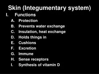
Functions of the Skin • Maintain homeostasis • Protective covering • Contains immune cells • Synthesizes vitamin D • Excretes wastes • Slows water loss • Regulates body temp • Houses sensory receptors
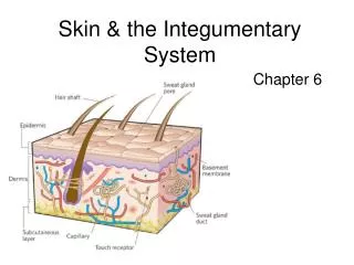
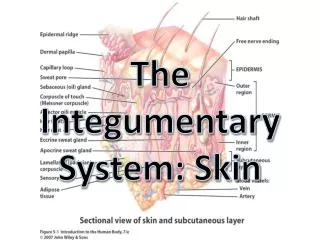
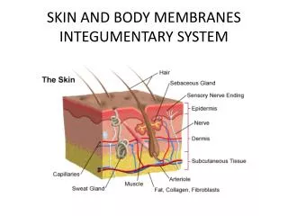
Cut-, skin Derm-, skin Epi-, upon Hypo-, below Layers of Skin • 3 layers: • Epidermis • Outer layer • Dermis • Inner layer • Subcutaneous • Hypodermis layer
Epidermis • Thickest on palms and soles • 0.8-1.4mm • Accessory structures • Hair follicles • Nails • Melanocytes • Provide melanin
Epidermis • Composed of stratified squamous epithelium • Keratinized • Cells synthesize keratin packed cells • Separated from dermis by basement membrane • Lacks blood supply
Layers of Epidermis • Stratum corneum • corneum= horn (Latin) • Outer keratinized layer, dead • Desquamation • Stratum lucidum • lucid= clear (Latin) • Palms, soles • Stratum granulosum • granulose= granular (Latin) • Flat, granular (keratohyalin)
Layers of Epidermis • Stratum spinosum • Spiny, prickly • Dividing, melanocyte branches • Stratum basale (germinativum) • Basal = basement, bottom layer • Melanocytes, dividing, deepest layer
Dermis • Bind epidermis to underlying tissue • Irregular dense CT • Muscle fibers • Smooth & skeletal • Dermal papillae • Increased surface area to nourish epidermis • Fingerprints • Friction for gripping • Average thickness • 1.0-2.0mm
Dermis • Accessory structures • Nerve cell processes • Pacinian (heavy touch/pressure) • Meissner’s (light touch/pressure) • Free nerve endings (temperature, pain) • Blood vessels • Sweat glands
Subcutaneous Layer • “Hypodermis” • Loose CT & adipose tissue • Major blood vessels • Insulation
Hair Follicles • From epidermal cells • Extends into dermis • Contains hair root • Hair color • Melanin production • Albinism • Arrectorpili muscle • Smooth muscle • Attaches to each follicle • Autonomic innervation
Hair Growth • Cyclic growth • Formed by cells in follicle • Keratinize and die • Form hair shaft • Type of hair • Eyelash • Grow 30 days, 100 days rest • Scalp • Grow 3-6 years, rest few months • 0.3 mm/day
Sebaceous Glands Seb-, grease • Associate with hair follicles • Absent on palms and soles • Holocrineglands • Release entire cells • Secrete sebum • Mixture of fatty acids and cellular debris • Secreted into hair follicle • Excess sebum acne
Clogged gland Excess sebum, epithelial cells Blackheads & whiteheads Good environment for anaerobic bacteria Clinical Application • Acne • Disorder of sebaceous glands • Hormonally influenced • At puberty • Adrenals androgens • Stimulates sebum production
Clinical Application • Immune system triggers inflammation • Pimple • Treatment options • Antibiotics • Estrogen • Retinoic acid (Vitamin A derivatives) • Accutane
Nails • Protective coverings on fingers, toes • Made of: • Nail plate • Nail bed (skin) • Lunula • White region • Half-moon shape • Active in growth • Reflection of health status • Blue = cyanosis; white = anemia; pigment = injury or melanoma; depressions/furrows = anemia, malnourishment; red streaks = ulcers, hypertension, RA • Growth rate • 0.5-1.2 mm/day
Sweat Glands • Sudoriferous glands • Widespread in skin • Deep dermis or subcutaneous layer • Eccrine glands • Most numerous • Apocrine glands • Active in puberty • Modified sweat glands • Ceruminous • Mammary
Sweat Glands • Eccrine glands • Most numerous • Response to • High temp • Exercise • Stress (on hands) • Open as pore • Forehead, neck, back
Sweat Glands • Apocrine glands • Onset @ puberty • Response to • Fear • Pain • Distress • Open into hair follicle • Armpit • Groin • Odor • Bacterial metabolism of secretions Sympathetic response
Sweat • Water • Salts • Sodium • Chloride • Potassium • Magnesium • Wastes • Urea • Lactate
Regulation of Body Temp • Heat • Product of cellular metabolism • Active cells are major heat producers • Skeletal muscle • As body temp , body releases heat • Homeostasis • Maintain body temp at 37C or 98.6F
Releasing Body Heat • Radiation (2)* • Most of heat loss • Infrared heat rays escape from warmer to cooler surroundings • Evaporation (5)* • At high temp, eccrine sweat glands release sweat onto skin • Heat carried away as skin cools • Conduction • Heat transferred from body directly to cooler object • Ex. Cold car seats • Convection (4) • Warm air circulates away from body
Conserving Body Heat • Dermal blood vessels • Reduce heat-carrying blood thru skin • Muscle activity • Increased cell respiration • Heat production • Shivering • Small groups of muscles contract • Produce heat
Problems in Body Temp Regulation • Hyperthermia • Abnormally high • Humid air prevents evaporation of sweat • No cooling • High air temp reduces radiation cooling • Hypothermia • Abnormally low • Shivering mental confusion, lethargy, loss of reflexes, shut down of major organs • Some surgeries (heart, brain) require body to be cooled less oxygen is required
Skin Color • Genetic Factors • Melanocytes • Same # in all people • Melanin production • Varies by person • Melanin production • High darker skin • Low fairer skin • Albinism • No melanin • Environmental factors • Darken existing melanin • Stimulate production • Sunlight • UV light from • Sunlamps • X rays • Tans fade • Pigmented epidermal cells keratinize • Wear away
Skin Color • Physiological Factors • Blood vessels in dermis adds color • Content of vessels • High oxygen hemoglobin is bright red • Pinkish hue to skin • Low oxygen hemoglobin is dark red • Bluish hue to skin (cyanosis) • State of vessels • Dilation skin reddens • Constriction skin pales • Dietary influences • Carotene yellow/orange skin tone • Health influences • Jaundice yellowish skin (liver)
Healing of Wounds and Burns • Inflammation • Response to injury or stress • Healing events depend on nature of injury • Cuts • Shallow • Epithelial cells divide fill in gap • Deep cut • Blood vessels break blood clot
Healing of Wounds and Burns • Clot • Scab formation • Fibrin, blood cells, platelets, fluids • Fibroblasts • Secrete collagen • Binds wound together • Scar • Extensive wound • Connective tissue on skin surface
Burns • First degree • Superficial partial thickness • Injuring epidermis only • Healing in 2-3 days • Common examples • Sunburn • Scalding water • Chemicals • Treatment • Flush with cool water (no ice) • Aloe (no oil) • Clean, dry bandage
Burns • Second degree • Deep partial thickness • Injures epidermis and dermis • Fluid escapes capillaries blisters • Healing time 1-2 weeks • Common examples • Prolonged sunburn or scalding water • Brief exposure to flame • Treatment • Flush with water, bandage • Do NOT break blisters • > 2-3 inches, see physician • Hydration • Antibiotics • Grafting
Burns • Third degree • Full-thickness • Epidermis, dermis, accessory organs • Healing time: weeks to months • Common examples • Contact with flame • Corrosive chemicals • Immersion in hot liquids • Treatment • Burn center • Debridement • Grafting
Grafting • Autograft • “Auto” self • Remove skin from unburned part of body and “transplant” it to injured site • Homograft • “Homo” like • Cadaveric skin used if can’t do autograft • Skin substitutes • Amniotic membrane • Artificial membrane • Cultured epithelial cells • Scarring
Healing of Burns • Treatment of patient: • Requires estimate of body surface injury • Replace body fluids and electrolytes • Determine amount of skin needed for graft • Use “rule of nines” • Divide skin surface into regions • Each region = 9% (or multiple of 9%)
Life Span ChangesAging skin shows many signs… • Cell cycle slows • Age (liver) spots • Dermis reduced • Connective tissue growth slows • Slower wound healing • Loss of fat • Wrinkles & sagging skin • Melanin production slows • Hair grays/whites • Less vitamin D production • Needed for calcium uptake in bones
Life Span ChangesAging skin shows many signs… • Hair growth slow • Thins and # follicles decreases • Lowered blood supply to nail beds • Dulls/hardens nails • Sensory receptors decline • Less sensitive to pain/pressure • Inability to control body temperature • # sweat glands drop & dermis blood vessel numbers
Common Skin Disorders • Athlete’s foot • Skin fungus infection (Tineapedis) • Boil • Bacterial infection, bacteria enter skin via follicle • Chickenpox • Varicella-zoster infection; blistery lesions that scab
Common Skin Disorders • Eczema • Dry, itchy, scaly skin (genetic) • Mole • Benign skin tumor (nevus) usually pigmented brown black • Psoriasis • Red skin w/ silvery scale
Rashes • Infectious • Roseola • Measles • Rubella • 5th disease • Shingles • Impetigo • Lyme disease • RMSF • Meningitis • Candidiasis • Allergic • Hives • Penicillin • Food allergies • Poison ivy • Cosmetics • Autoimmune • Lupus • Psoriasis
Skin CancerTypes • Basal cell carcinoma • Most common • Basal layer of epidermis • Nodule, shiny bump, scar-like lesion • Squamous cell carcinoma • 2nd most common • Upper layers of skin (squamous) • Begin as scaly red patches, open sores, elevated with centralized depression • Typically superficial
Skin CancerTypes • Malignant melanoma • Most deadly • Brown/black patches, nodules • Look like or arise from moles
Skin CancerRisks • Fair skin, hair, eye color • Family history • Personal history • Chronic sun exposure • History of sunburns early in life • Certain types of moles, # moles • Freckles • Sun sensitivity, sun damage
Skin CancerDetection • A • Asymmetry • B • Border irregularity (scalloped, notched) • C • Color variation • D • Diameter > ¼ inch • E • Evolution
Skin CancerTreatment • Excision • Virtually all types of skin cancer are 100% curable if caught early • Invasive • Lymph node testing • Chemotherapy • Immunotherapy
Suggested Homework Problems • Chapter assessments • 3-6, 9, 11-14, 17, 19-25, 27, 28 • Integrative Assessments/Critical Thinking • 1-7