Download
1 / 51
530 likes | 730 Vues
Acute respiratory distress syndrome. ALOK SINHA Department of Medicine Manipal College of Medical Sciences Pokhara , Nepal. alveolar epithelium. Serious disease characterised by damage to alveolar epithelium & capillary endothelium resulting in alveolar oedema with high protein fluid

E N D
Acute respiratory distress syndrome ALOK SINHA Department of Medicine Manipal College of Medical Sciences Pokhara, Nepal
alveolar epithelium • Serious disease characterised by damage to alveolar epithelium & capillary endothelium resulting in alveolar oedema with high protein fluid • It results from increased alveolar capillary permeability • NOT CARDIOGENIC IN ORIGIN Capillary endothelium
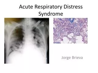
severe end of a spectrum of acute lung injury due to many different insults. Essentially it is diffuse alveolar injury • Acute, persistent, lung inflammation • Increased vascular permeability • Bilateral and extensive infiltrates (seenin the CXR) • Very poor oxygenation despite PEEP
Not due to clinical left ventricular failure – associated with a wedge pressure of over 18 mmHg (non-cardiogenic) • Most commonly seen on the ICU where about 10% of such patients will have ARDS
PCWP Normal: 8-12mmHg
International criteria 1. Acute onset of symptoms 2. PaO2 : FIO2(fraction of inspired oxygen) – 200 mm Hg or less For Example normal PaO2 = 100, FiO2 = 20% or 1/5 so the above ratio = 100/1/5 = 500 mm Hg 3. Bilateral infiltrates on CXRs 4. Pulmonary arterial wedge pressure of 18 mm Hg or less (or no clinical signs of left atrial hypertension)
What is this situation ? 1. Acute onset of symptoms 2. PaO2 : FIO2: between 300 to 200 mm Hg 3. Bilateral infiltrates on CXRs 4. Pulmonary arterial wedge pressure of 18 mm Hg or less or no clinical signs of left atrial hypertension • Acute lung injury (ALI) Mild ALI ARDS
Inflammatory damage to the alveoli • by locally produced pro-inflammatory mediators • remotely produced and arriving via • pulmonary artery • Through inhalation (eg gastric contents) • Increased pulmonary capillary permeability - fluid and protein leakage into the alveolar spaces with pulmonary infiltrates • Alveolar surfactant is diluted with loss of its stabilizing effect, resulting in diffuse alveolar collapse and stiff lungs. This leads to:
1. Gross impairment of V/Q matching with shunting causing arterial hypoxia • usually enough remaining functioning alveoli to maintain CO2 clearance- Normal CO2 • Pulmonary hypertension will develop secondary to the hypoxia (helpful – counters V/Q matching) 2.Reduced compliance (stiff lungs), due to loss of functioning alveoli • alveolar collapse, filled with fluid and protein • hyperinflation of remaining alveoli to their limits of distension Decrease transfer of gases in alveoli 3.
Causes of mediator release leading to ARDS • Sepsis/pneumonia • Gastric aspiration • Major trauma • Smoke/ gas inhalation • Acute pancreatitis • Drug toxicity - tricyclic antidepressants, opiates, cocaine, aspirin • Fat emboli • Direct effects of large amounts of necrotic tissue (secondary risk factors) • alcoholism • cigarette smoking
Less common causes • Near drowning • Following upper airway obstruction: mechanism unclear • Acute form of Interstitial Pneumonia: Also known as acute Hamman-Rich syndrome • Post-bone marrow transplant as bone marrow recovers • Amniotic embolism • Massive haemorrhage • Multiple transfusions • DIC
Massive burns Head injury Raised ICP Intracranial bleed Cardio-pulmonary bypass Acute liver failure
Phase 1 - early period of alveolar damage and hypoxaemia with pulmonary infiltration • Phase 2 -develops after a week as pulmonary infiltrates resolve • associated with an increase in • type II pneumocytes (surfactant producers) • Myofibroblasts • collagen formation
Phase 3 -if the patient survives, is the fibrotic stage that leaves the lung with • Cysts • deranged micro-architecture • fibrosis on histology • leading to Cor Pulmonale
CLINICAL FEATURES
ARDS should be considered in any patient with a predisposing risk factor • develops severe hypoxaemia • stiff lungs • widespread diffuse pulmonary infiltrate Approximately 1 to 2 days following the clinical presentation of the precipitating cause
Rapidly worsening dyspnoea • Dry cough • Hypoxaemia • Coarse crackles in the chest
DIFFERENTIALS Aspiration Pneumonia Congestive Heart Failure Pneumonia Atypical Bacterial Pneumonia Pneumocystis Carinii Pneumonia Viral
To exclude other more specifically treatable conditions • Left ventricular failure excluded • on clinical grounds • by echocardiography • wedge pressure measurement <18 mmHg.
Diffuse alveolar haemorrhage can occur in Goodpasture's, Leptospirosis Oher clinical features of these disorders will be present • Some pulmonary infections • Mycobacteria • Legionella • PCP • viral pneumonia may mimic ARDS & lavage fluid may reveal these • Occasionally cancer and lymphangitis carcinomatosa can also mimic ARDS • will show on a lung biopsy
CXR • ABG (consider arterial line as regular samples may be required) • CBC,LFTs, coagulation profile, and CRP • Septic screen (culture blood, urine, sputum) • ECG • Consider drug screen • Amylase if history suggestive
Pulmonary artery catheter to measure • PCWP • cardiac output • mixed venous oxygen saturation • calculation of haemodynamic parameters • Other investigations if appropriate • CT chest • Broncho alveolar lavage for microbiology & cell count (?eosinophils)
Treat the precipitating cause • Provide best supportive care with adequate oxygenation • I.V. fluids – • to be used judiciously • Can increase pulmonary oedema
Inotrope and/or vasopressor support is commonly required and the choice of agent is • dobutamine • dopamine • epinephrine, norepinepherine • Patients invariably require higher oxygen concentrations (non-rebreather masks with reservoir FiO2 ~60- 80%) or CPAP • Consider transfer to HDU/ICU
Different oxygen delivery systems Nasal canula Non re breather masks
Indications for mechanical ventilation • Inadequate oxygenation (PaO2 <60mm (8kPa) on FiO2 >0.6 or 60%) • Rising or elevated PaCO2 (> 45 mm or 6kPa) • Clinical signs of incipient respiratory/cardiovascular failure
Mechanical ventilation with PEEP -almost always required to maintain oxygenation, with high inflation pressures Is this the meaning of PEEP ? PEEP: required to counter atelectasis
High inflation pressures may worsen ARDS directly (micro-barotrauma) • try to maintain plateau pressures <30 mmHg
Special ventilation techniques have been tried to reduce the high inflation pressures resulting from the stiff lungs (low compliance) • Using low tidal volumes to reduce inflation pressures (6 ml/kg ideal body weight compared to 12 ml/kg) reduces mortality by 10% • This results in • Reduced minute ventilation • Rise in PaCO2 – permissive hypercapnia
Inverse ratio ventilation • may improve oxygenation, but pCO2 may rise further • Prone positioning • improves oxygenation in ~70% of patients with ARDS
Inhaled pulmonary vasodilators (nitric oxide, nebulized prostacyclin): may improve oxygenation Extracorporeal oxygenation/CO2 removal will buy time and allow the lung to recover, but these techniques are very expensive and it is difficult to demonstrate any long-term benefit
High-dose steroids – • some evidence of overall improved survival • later use possibly beneficial if nosocomial infection rates are not increased
Cardiovascular support Most patients haemodynamically compromised due to • underlying condition • ventilatory management • Benefit from fluid resuscitation. This may risk worsening capillary leak in the lung and compromise oxygenation/ventilation. Aim for a low-normal intravascular volume whilst maintaining cardiac index and mean arterial pressure
Management of other associated conditions • Renal failure • Enteral feeding • Coagulopathy -severe/DIC may be present expert advice should be sought • Sepsis • empiric antibiotics guided by possible pathogens, and following an appropriate sensitivity tests Antibiotics should be modified or discontinued in light of microbiological results
High ventilation pressures lead to barotrauma: • pneumothorax • surgical emphysema • pneumomediastinum • Nosocomial infections • Non-specific problems of • venous thromboembolism • GI haemorrhage • inadequate nutrition
Prognosis • has improved over the last 20 years due to improvements in supportive care of • Early deaths due to the precipitating condition • later deaths to complications • Over half the patients will survive with varying residual lung damage, pulmonary function tests often show only minor restrictive abnormalities
Future developments • The optimal level of PEEP is difficult to predict. • Inadequate PEEP allows more atelectasis • too high PEEP contributes to overdistension of remaining alveoli and further barotrauma • Ways to estimate the best PEEP are under investigation
Liquid ventilation with perfluorocarbons has been tried • Nitric oxide (NO) has been tried with clear improvements in oxygenation but very little effect on survival • Inhaled prostacyclin: is unconvincing
bilateral patchy opacities in mostly the middle and lower lung zones
Normal size heart No pleural effusion