Download
1 / 23
260 likes | 787 Vues
COPD. Michele Ritter, M.D. Argy Teaching Resident, Feb. 2007. Definition .

E N D
COPD Michele Ritter, M.D. Argy Teaching Resident, Feb. 2007
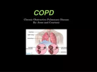
Definition • A disease state characterized by the presence of airflow obstruction due to chronic bronchitis or emphysema; the airflow obstruction is generally progressive, may be accompanied by airflow hyperactivity, and may be viewed as partially reversible. • Includes emphysema and chronic bronchitis
Prevalence • COPD occurs in 4-6% of white males, and 1-3% of adult white females • The 4th most common cause of death in the United States • 14.2 million people in U.S. have COPD • Highest mortality rate is in white men, and the lowest is in hispanic women.
Types of COPD • Emphysema • Permanent and destructive enlargement of airspaces distal to the terminal bronchioles without obvious fibrosis and with loss of normal architecture • Always involves clinically significant airflow limitation. • “pink puffer” • Chronic Bronchitis • Presence of a cough productive of sputum not attributable to other causes on most days for at least 3 months over 2 consecutive years • May be present in the absence of airflow limitation. • “blue bloater”
Pathogenesis of COPD • Increased number of activated polymorphonuclear cells and macrophages produce elastases (such as human leukocyte elastase), resulting in lung destruction. • Increased oxidative stress caused by free radicals in cigarette smoke, the oxidants released by phagocytes, and polymorphonuclear leukocytes all may lead to apoptosis or necrosis of exposed cells
Emphysema 3 morphologic patterns: Centricacinar: focal destruction limited to the respiratory bronchioles and the central portions of acinus associated with cigarette smoking most severe in the upper lobes Panacinar: involves the entire alveolus distal to the terminal bronchiole develops in patients with homozygous alpha1-antitrypsin (AAT) deficiency most severe in the lower lung zones Distal acinar: Also called paraseptal least common form involves distal airway structures, alveolar ducts, and sacs localized to fibrous septa or to the pleura and leads to formation of bullae (can result in pneumothorax) Chronic Bronchitis Mucus gland enlargement Airway atrophy, focal squamous metaplasia, ciliary abnormalities, variable amounts of airway smooth muscle hyperplasia, inflammation, and bronchial wall thickening Respiratory bronchioles display a mononuclear inflammatory process, lumen occlusion by mucous plugging, goblet cell metaplasia, smooth muscle hyperplasia, and distortion due to fibrosis Airway walls to deform and narrow the airway lumen Pathogenesis of COPD (cont.)
Risk Factors • SMOKING! • 48 million smokers in the U.S. • 3000 new people take up smoking daily • Nearly all patients with symptomatic COPD are current or former smokers • 10-20% of smokers will develop symptomatic COPD. • In men who smoke one pack/day, the drop in FEV1 per year was 9 mL more than in non-smokers • Occupational Exposures • Dusts, gases, fumes • Alpha1-antitrypsin deficiency • Alpha1-antitrypsin is an important protease inhibitor that usually presents elastases from causing lung destruction
Symptoms • Dyspnea • Cough (usually worse in morning, sputum production) • Wheezing • Cyanosis • Right heart failure • Weight loss, anorexia
Physical Exam • RR, HR, O2 saturation • Gen: Barrel-chest, accessory muscle use • CV: Quiet heart sounds • Resp: Decreased breath sounds, wheezing, rhonchi, crackles
Labs • CBC: Hgb/Hct • ABG: pH, pCO2 • Chemistry: HCO3
Diagnosis of COPD • Look for secondary polycythemia: • Hct >52% in males, Hct>47% in females • Measure alpha1-antitrypsin levels in all patients 40 years or younger, or in those with family history. • Hyperinflation see on chest x-ray • Bullae seen on Chest x-ray or CT scan
Diagnosis of COPD – Pulmonary Function Tests • Forced Expiratory Volume for 1 second (FEV1) • FEV1/FVC (Forced Vital Capacity) ratio • Total Lung Capacity (TLC) • Forced Residual Capacity (FRC) • Residual Volume (RV) • Vital Capacity (VC)
COPD Exacerbation • Typically manifest as increased sputum production, more purulent sputum and worsening of dyspnea. • Although infectious etiologies account for most exacerbations, exposure to allergens, pollutants or inhaled irritants may also play a role. • Bacterial infection is a factor in 70 to 75 percent of exacerbations, with up to 60 percent caused by • Streptococcus pneumoniae • Haemophilus influenzae • Moraxella catarrhalis • Antibiotic therapy has a small but important effect on clinical recovery and outcome. • Respiratory fluoroquinolone (Levofloxacin, Moxifloxacin) • Ceftriaxone + azithromycin • Short courses of systemic corticosteroids may provide important benefits in patients with exacerbations of COPD. • Oxygen therapy to keep saturation Between 90-93% • Non-invasive ventilation such as BiPAP can be helpful in avoiding intubation/mechanical ventilation.
Treatment of COPD • SMOKING CESSATION! • Short-acting bronchodilators • albuterol • Long-acting bronchodilator • salmeterol • Combination of anti-cholinergic and -agonist bronchodilator • Ipratropium + albuterol (combivent) • Tiotropium (spiriva) • Methylxanthines (Theophylline) • Has anti-inflammatory affect, and improves respiratory muscle function, stimulates the respiratory center, and promotes bronchodilation • Adverse effects include anxiety, tremors, insomnia, nausea, cardiac arrhythmia, and seizures • Inhaled corticosteroids • Fluticasone (Flovent), budesonide (Pulmicort) • Combination of Inhaled corticosteroid and long-acting -agonist • Fluticasone + salmeterol (Advair) • Oral Corticosteroids
Treatment of COPD (cont.) • Oxygen Therapy • Continous oxygen has been shown to cut mortality in half or decrease morbidity when compared with non-continous oxygen • Continuous (24 hours/day) • Resting Pa02 of 55 mm HG, or Resting oxygen saturation < 88% • Resting Pa02 of 56-59 mmHg or Oxygen Sat. <89% in presence of dependent edema (suggestive of CHF), P pulmonale on ECG (P wave more than 3 mm in inferior leads) or cor pulmonale, or erythrocytosis (Hct > 56) • Noncontinuous • During exercise – when PaO2 is < 55 mmHg or Oxygen sat. < 88% with low level of exercise. • During sleep if Pa02 is < 55 mmHg or Sa02 less than 88% with associated complications such as pulmonary hypertension, daytime somnolence, cardiac arrythmias.
Treatment of COPD (cont.) • Pulmonary Rehabilitation • Aimed at keeping patient conditioned with exercise, perception of dyspnea, quality of life and self-efficacy. • Surgery • Bullectomy • Resection of large bullae compressing normal lung • Lung volume reduction surgery • Pneumonectomy of nonuniform emphysematous lung • Double lung transplantation • Can be life-saving, but is costly, can be lack of donor availability and requires lifelong immunosuppression.
Take Home Points • Smoking is the number one cause of COPD! • If smoking is stopped once COPD diagnosed, the progression of disease can slow down. • Treat COPD exacerbations with antibiotics and possibly with steroids. • Continuous oxygen is shown to decrease morbidity and mortality in COPD