Download
1 / 35
350 likes | 466 Vues
P1-1 Cell diagram . 3P 1-1. Chromatin to DNA. From chromosome to chromatin to solenoid to DNA . P3-2 Chromosome to DNA . 3P3-2. Chromosomes. P3-2 Backbone chemistry. 5. 3. 5. O. 4. 1. 3. 2. 3P3-2. 4F4-4. 4F4-3. 3F2-10. 8-30 chromatin. 3F8-30. Tangle of chromatin spilling

E N D
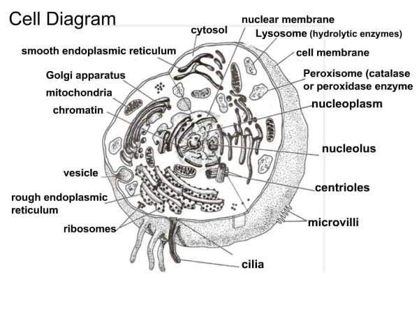
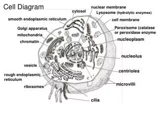
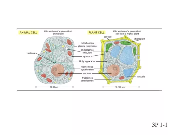
P1-1 Cell diagram 3P 1-1
Chromatin to DNA From chromosome to chromatin to solenoid to DNA
P3-2 Chromosome to DNA 3P3-2 Chromosomes
P3-2 Backbone chemistry 5 3 5 O 4 1 3 2 3P3-2
4F4-3 3F2-10
8-30 chromatin 3F8-30
Tangle of chromatin spilling out of lysed nucleus 2 linked copies of a chromosome during mitosis 4F4-21
Transcription Translation mRNA Protein Gene (DNA) Central Dogma Transcription Reverse Transcription
Gene Gene 27Kbp 27Kbp 300Kbp 300Kbp Intron1 Ex1 Ex2 Intron2 145bp 2500bp 145bp 2500bp 5’ UTR 3’ UTR JUNK???
3-12,13 DNA replication Template, 3’ New, 5’ 3’ 5’ DNA Synthesis 4F5-3 4F5-5
proteins- chain of AA NH3+ NH NH NH O O O O C C C C C C C C H H H H R2 R1 R3 R4 Peptide bond R2 R1 R2 O -- R1
Peptide bond,crambin CRAMBIN: N = 46 TTCCPSIVARSNFNVCRLPGTPEALCATYTGCIIIPGATCPGDYAN
4F6-9 3F3-19 4F6-8
Transcription Start Site TATA G9.17
The Gene Network of E.Coli: (bacterium, single cell) (U. Alon)
M.Milyevsky,Y.Tabach, ….T.Pilpel,V. Rotter – in vitro • 2.C. Rosty,M. Sheffer, D.Tsafrir….in vivo, cervical cancer Degradation Induced by E6 Inactivation by E7 binding p53 CDKN1A (p21) CDK2 NFY CHR RB NFY CDK4 CDK6 CDKN2A (p16) E2F CDE / E2F ELK1 Mitogens MAPK ELK 1 Cervical cancer induced by HPV: two viral oncoproteins (E6,E7) are inserted in DNA and expressed. They inactivate p53 and Rb and induce proliferation
3-16 Genetic code 3 READING FRAMES
beta-globin(human)sequences VHLTP(EEKSAVTALW){GK} VN(VDEVGGEALGRLLVV)Y (PWTQRF)F(ESF)GDLST (PDAVM){G}N (PKVKAHGKKVLGAFSDGL) {AH}(LDNLKGTFATLSELHCD) {KL}HVD{P} (ENFRLLGNVLVCVLAHHFGKE)FT (PPVQAAYQKVVAGVANALA) {HK}YH
6-8,9 tRNA 6-8,9
4F6-65 3FtRNA
4F6-58 4F6-60
polyribosome 3F6-28,29
Transcription Translation mRNA Protein Gene (DNA) Central Dogma Cells express different subset of the genes In different tissues and under different conditions